
The complete 3D spatial biology platform
Aurora combines whole-tissue imaging, automated data management, and AI-powered analysis in a unified workflow for translational research and digital pathology.
Aurora workflow
How the Aurora 3D™ Spatial Biology Platform Works
The Aurora 3D™ Spatial Biology Solution is a unified platform that converts intact tissue into quantitative 3D datasets. It includes three integrated modules.
3Di™ is the proprietary light-sheet microscope for high-resolution 3D tissue imaging.
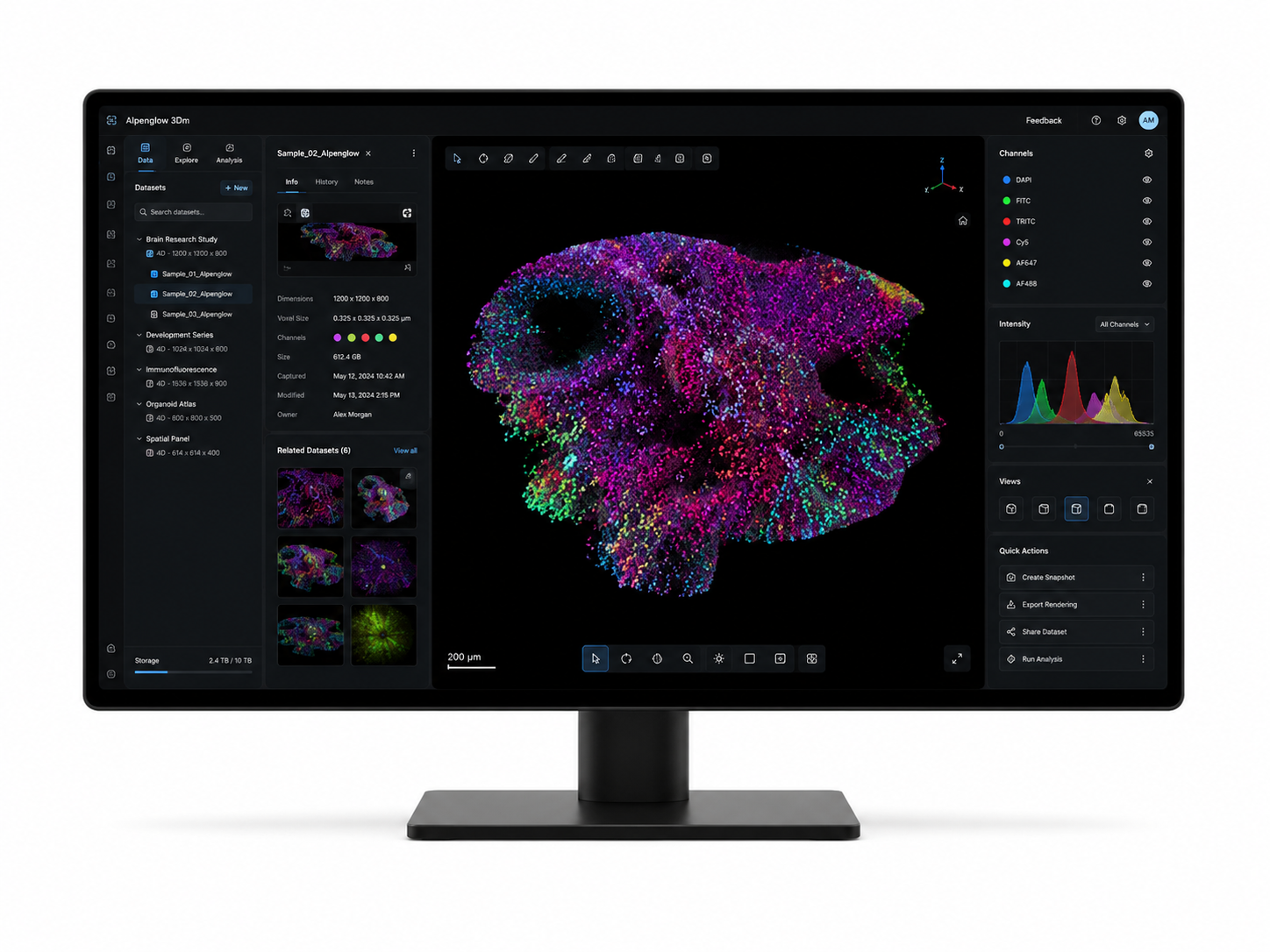
3Dm™ handles automated data management and processing for large volumes.
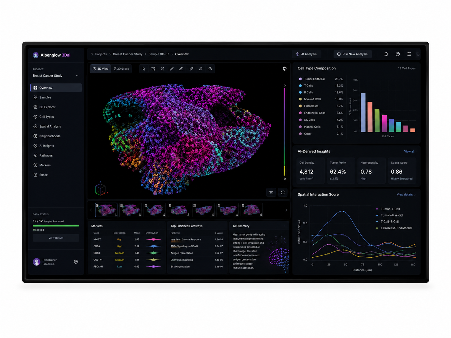
3Dai™ provides AI-powered image analysis for full tissue quantification.
Intact Tissue
3Di™
Imaging
3Dm™
Data Management
3Dai™
AI Analysis
Biological Insight
Aurora 3D™ Platform can be accessed in multiple ways.
The 3Di™ light-sheet microscope is available for purchase as a standalone instrument or can be combined with 3Dm™ and 3Dai™ to create an in-house workflow.
The whole platform is also available through Alpenglow’s 3D histology imaging services , providing complete imaging, processing, and analysis.
3D tissue imaging
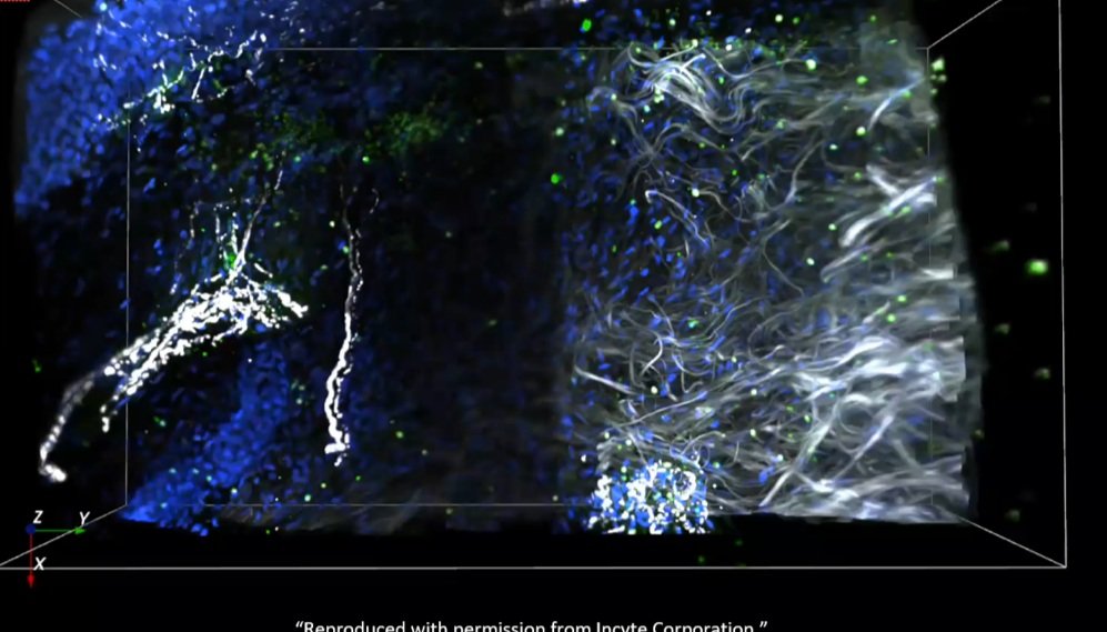
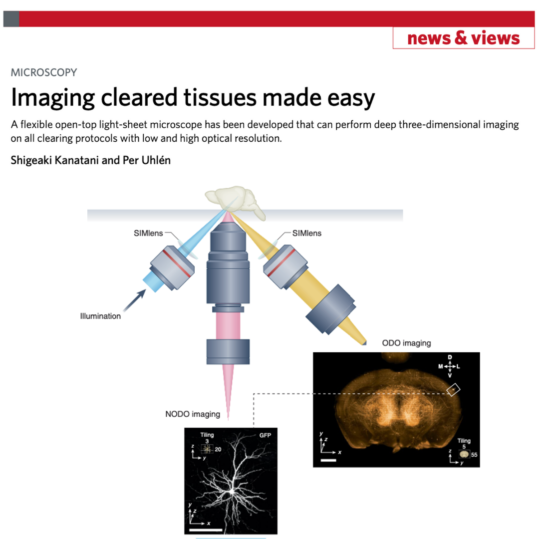
3D Tissue Imaging: 3Di™ HOTLS
High-resolution whole tissue imaging with Hybrid Open-Top Light-Sheet Microscopy
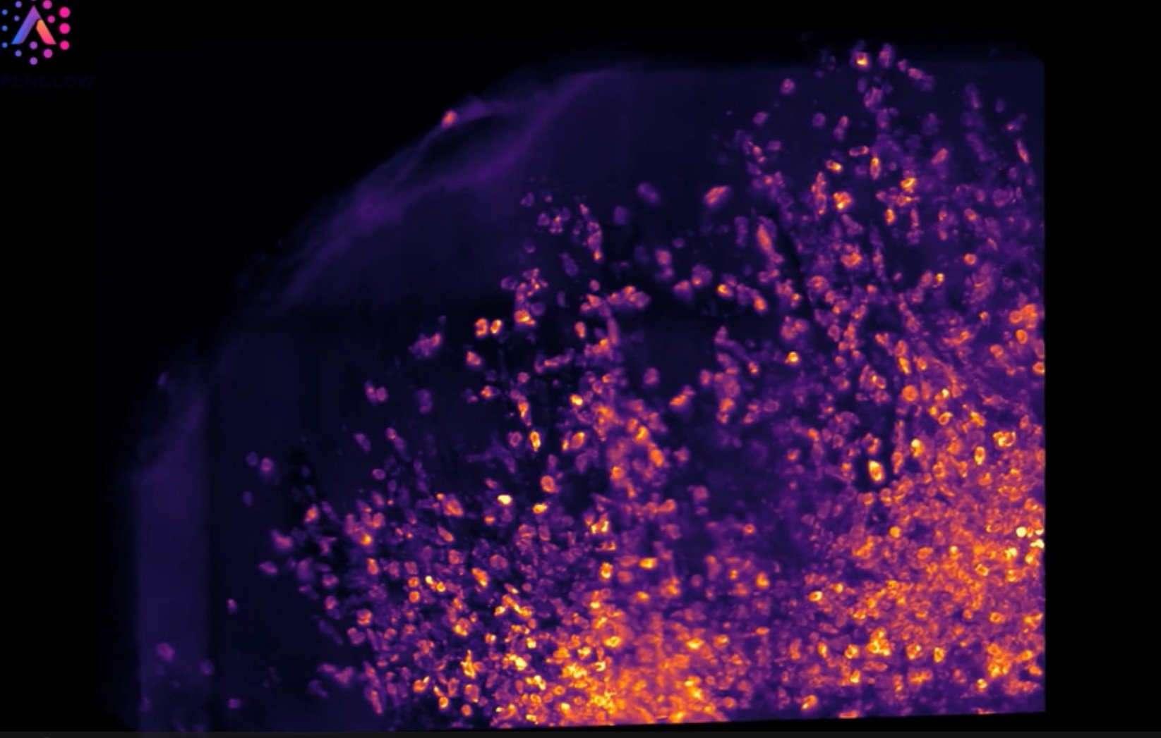
The Aurora 3D™ Platform includes the 3Di™ Hybrid Open-Top Light-Sheet (HOTLS) Fluorescent Microscope. It captures intact tissue blocks using structured illumination and multi-scale imaging, generating clean, high-resolution, submicrometer-detail volumetric datasets. The system supports a 1 cm working distance and scan dimensions of 12.5 cm × 7 cm, as described in the Nature Methods publication. It delivers non-destructive fluorescent imaging across skin biopsies, tumors, lymph nodes, organs, and preclinical models.
Data management
Data Management: 3Dm™
Automated registration, alignment, and flat field correction for large 3D data sets
3Dm™ organizes and processes large 3D tissue volumes generated by the 3Di™ system. It manages stitching, illumination correction, multi-channel integration, and volume optimization for fluorescent imaging data.
The pipeline is designed for fluorescent imaging data and supports multi-scale datasets that span scout and zoom acquisitions. This comprehensive tool automatically aligns images post-imaging, and is validated for data sizes exceeding 4 terabytes.
AI-powered analysis
AI-Powered Analysis: 3Dai™
Quantification, spatial profiling, and biological readouts from intact tissue
3Dai™ extracts biological meaning from whole tissue datasets using AI-powered analysis. It identifies cells and structures across the entire volume, measures spatial relationships, and provides 3D metrics that are not accessible from traditional 2D sections.
Analysis can include cell segmentation, immune profiling, nerve and vessel quantification, volumetric measurements, region-level stratification, and multi-channel spatial mapping. The platform delivers analysis-ready outputs that support biomarker discovery, translational research, computational modeling, and quantitative spatial biology workflows.
Why Aurora
One connected platform from tissue to insight
Aurora 3D™ combines whole tissue imaging, automated data management, and AI-powered analysis in one spatial biology workflow.
Separate tools
- Disconnected imaging and analysis
- Manual handoffs between steps
- Harder to standardize outputs
Connected platform
- Imaging, processing, and analysis together
- Automated volumetric data management
- Quantitative spatial biology readouts
Image tissue
Whole tissue imaging for biopsies, organs, tumors, and organoids.
Manage data
Align, organize, and process large 3D datasets.
Analyze biology
Quantify cells, structures, and spatial patterns.
Use results
Readouts for biomarkers, digital pathology, and translational research.
Aurora applications
Apply 3D spatial biology across tissue-driven research
Aurora 3D™ Platform supports programs where tissue architecture, cell distribution, and microenvironmental context are essential.

Workflow example
Watch Aurora turn intact tissue into measurable 3D data
This example follows a 3D heart vasculature workflow, from imaging to quantitative readouts such as cell counts, vessel dimensions, distance measurements, and branching patterns.