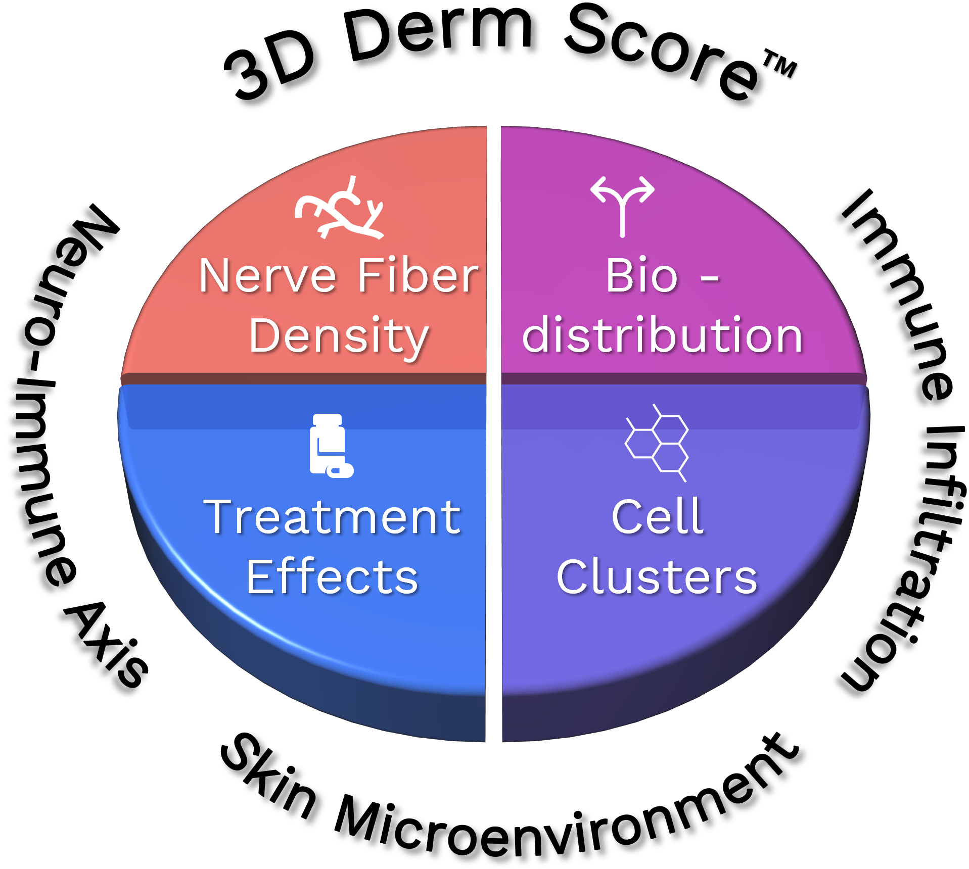
Quantifying Skin Architecture in Three Dimensions with the 3D Derm Score™ Assay.
3D Derm Score™ Assay:
Advanced Analysis of the Skin Microenvironment
The 3D Derm Score™ Assay is a cutting-edge, end-to-end solution designed to comprehensively analyze the skin microenvironment and the neuro-immune axis in skin diseases. Our Aurora platform offers an unprecedented level of detail, enabling an in-depth assessment of peripheral innervation and cutaneous inflammation.
Key Benefits
Comprehensive, high-resolution 3D imaging of the skin microenvironment, including detailed study of the Nerve Fiber Density.
Proprietary data analysis workflow for accurate and efficient results.
Flexible marker customization with a wide selection of pre-optimized options.
Support for diverse research needs across various diseases (e.g., Atopic Dermatitis, Hidradenitis Suppurativa, Alopecia, Prurigo Nodularis).
3D Derm Score™ Assay:
The Workflow
The complete workflow begins with 3D imaging using Alpenglow Biosciences’ advanced 3Di™ Hybrid Open-Top Light-Sheet Microscope, ensuring high-resolution imaging of skin samples. The generated 3D data is then seamlessly managed and analyzed through our proprietary software, 3Dm™, and 3Dai™, which deliver precise, data-driven insights tailored to your research objectives.
We provide flexibility and customization for marker selection.
Choose from a set of pre-optimized marker panels, or design your own by selecting from our established markers or having your preferred markers optimized.
Our platform supports easy marker swapping to accommodate the evolving needs of your experiments.
We offer optimized markers for multiple species, including humans and mice.
3D Derm Score™ Assay:
Markers selection
Option A
Select a pre-optimized panel
Option B
Design your panel
(Example markers shown)
Discover the Hidden Depths of Skin in 3D
3D Imaging of Scalp Tissue stained with TO-PRO-3 red for nuclei, CD45 blue for immune cells and PGP9.5 green for nerves. 3D imaging captures the epidermal layer, nerve distribution, and piliferous bulb structure in measurable detail.
High-resolution 3D imaging of a Region of Interest in a lesional Atopic Dermatitis sample, stained with TO-PRO-3 blue for nuclei, PGP9.5 white for nerves, and CD45 yellow for lymphocytes. Witness the intricate innervation within the epidermis and dermis and the precise identification of immune-cell clusters located near specific nerves.


