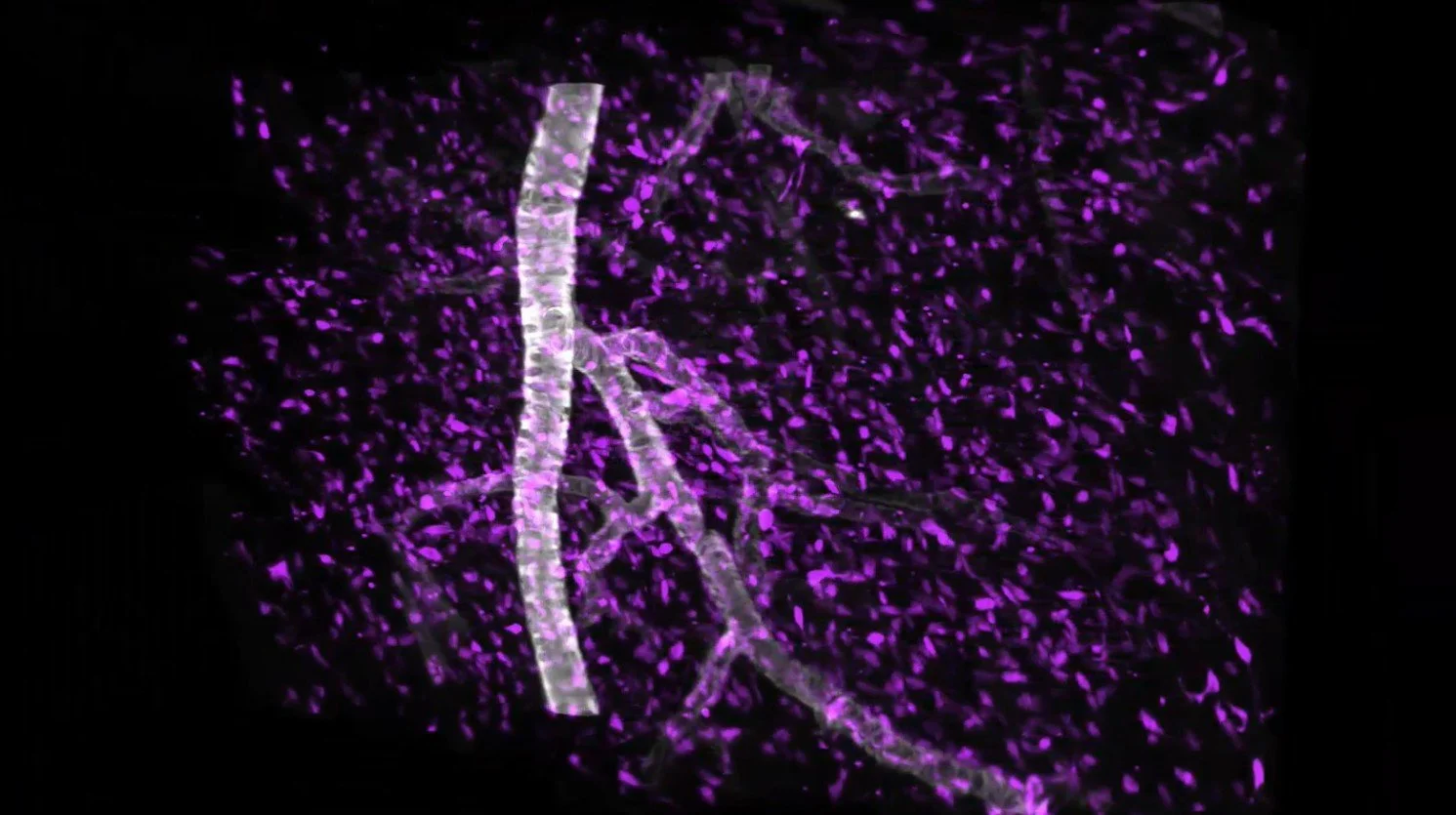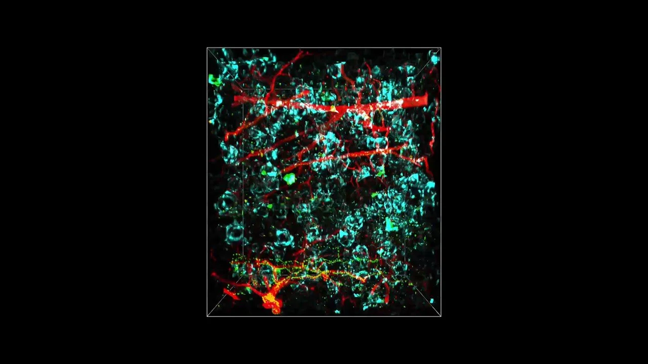
SEE ENOUGH OF THE TISSUE TO TRUST THE CONCLUSION
Turn intact tissue into quantitative 3D data
Alpenglow combines 3D light-sheet imaging, automated volumetric data processing, and AI-powered analysis to quantify cells, structures, and spatial relationships for drug development, diagnostics, and AI.
How to work with Alpenglow
Choose how to access 3D imaging
Outsourced 3D histology
Complete workflow performed by Alpenglow
Send tissue for end-to-end 3D analysis
Send fixed tissue to Alpenglow for clearing, staining, 3D light-sheet imaging, and quantitative analysis. Get whole-sample 3D data without building the workflow in-house.
In-house 3D imaging platform
Instrument operated within your facility
Bring 3D imaging into your facility
Install the 3Di™ Hybrid Open-Top Light-Sheet microscope and connected software to run whole-sample 3D imaging within your laboratory or shared facility.
Applications
See what whole-sample 3D can measure
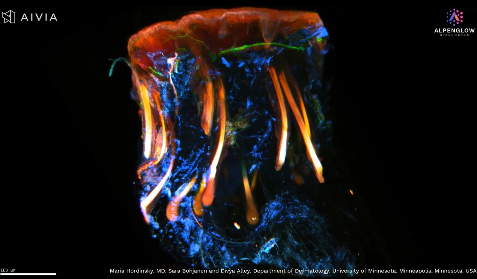
Dermatology
Map skin biology across whole biopsies
Follow nerves, immune cells, follicles, vessels, and epidermal architecture across intact skin biopsies.
Nerve density, immune distribution, follicular organization, spatial distances, and regional variation.
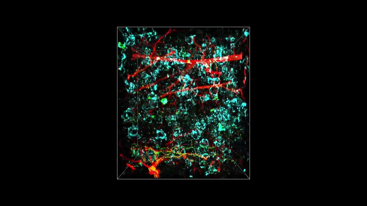
Immuno-oncology
Quantify immune organization across intact tumors
Map immune infiltration, exclusion, tumor boundaries, vasculature, collagen, and TLS across intact tumors.
Cell density, infiltration depth, clustering, TLS volume, spatial relationships, and tissue heterogeneity.

Multi-field capabilities
Analyze connected structures across tissues
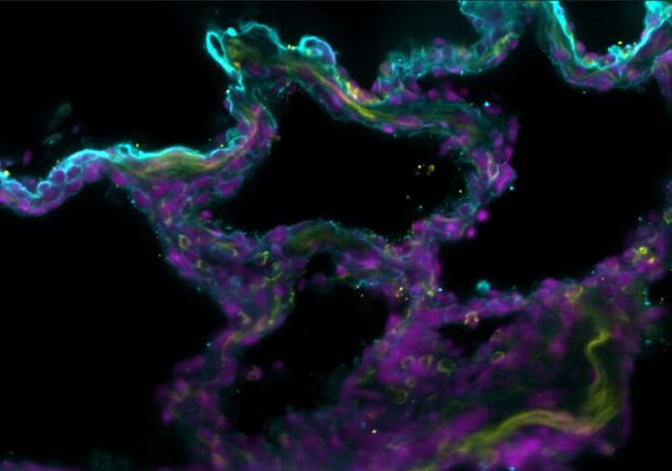
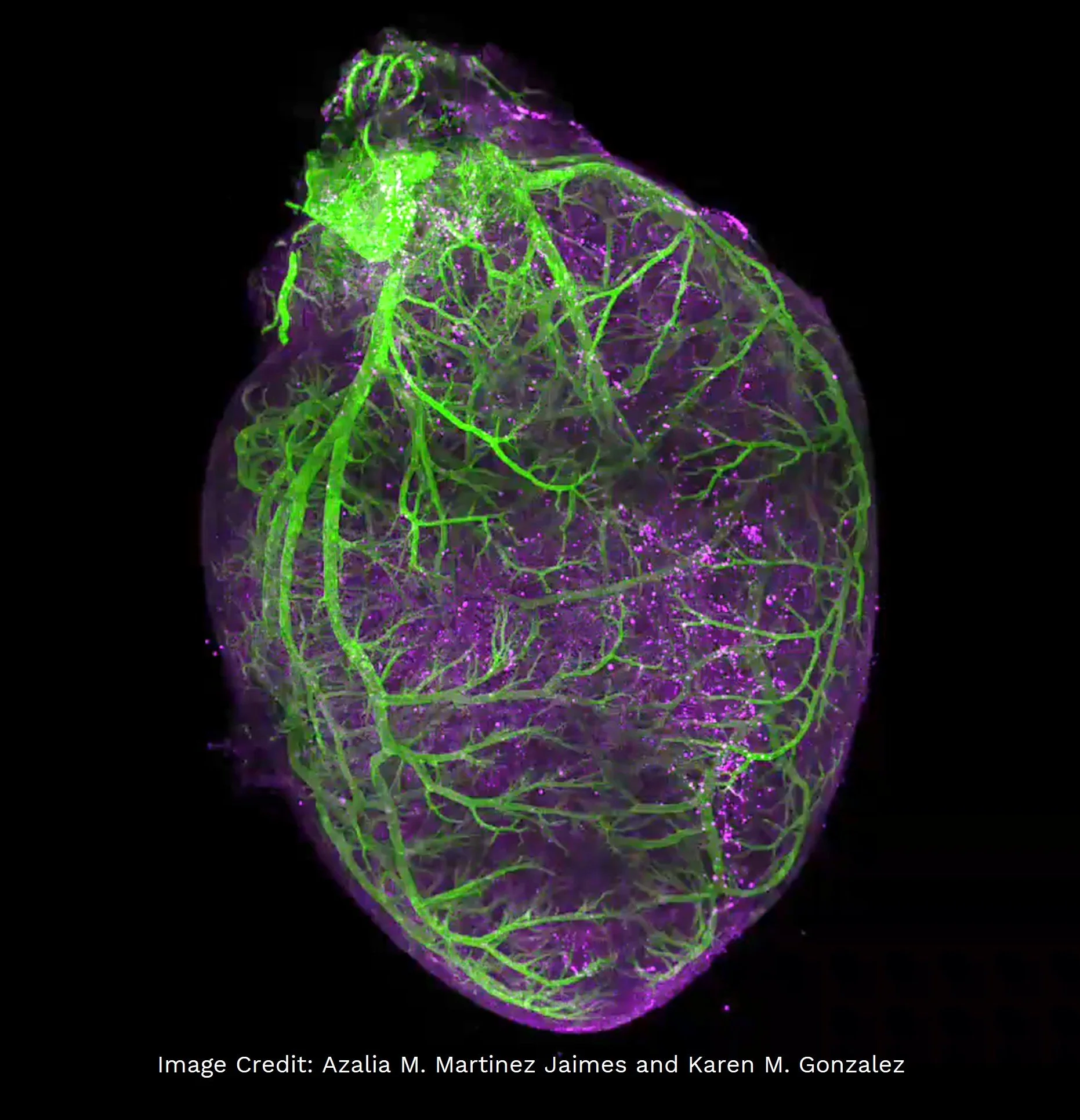
Trace villi, crypts, vessels, nerves, airways, ducts, and other connected structures across tissues and model systems.
Branching, diameter, volume, morphology, density, structural continuity, and regional distribution.
Aurora 3D™ Spatial Biology Solution
Capture intact tissue. Turn the volume into quantitative data.
Imaging hardware
Acquisition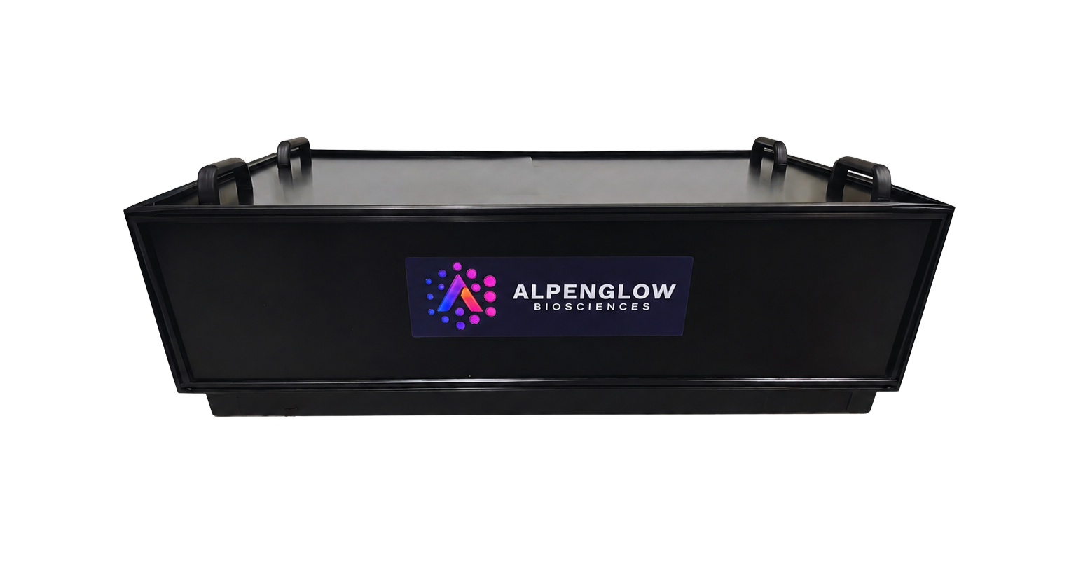
3Di™
Hybrid Open-Top Light-Sheet microscope
Image intact tissue
Capture whole-sample context and high-resolution regions of interest with a dual-path Scout-to-Zoom workflow.
ExploreData and analysis software
Processing and quantification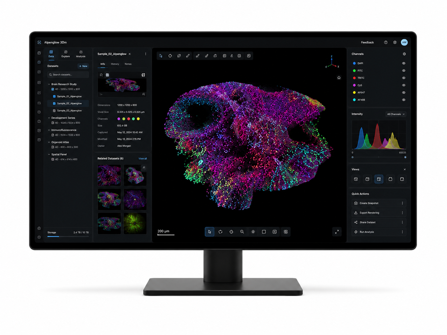
3Dm™
Volumetric data management
Process the volume
Process and organize multichannel 3D datasets for review and downstream analysis.
Explore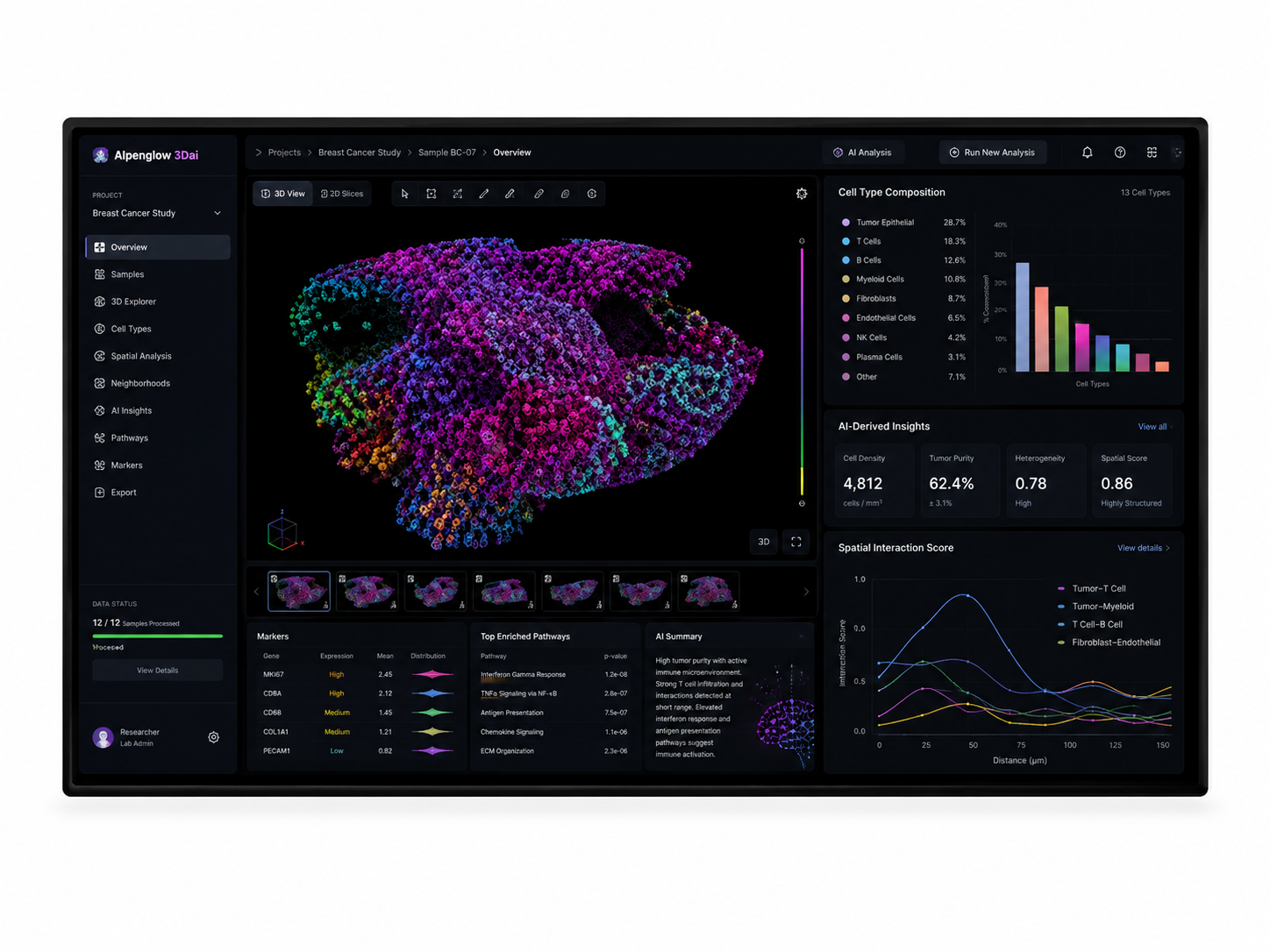
3Dai™
AI-powered image analysis
Quantify the biology
Segment cells and structures, measure spatial relationships, and quantify biology across intact tissue volumes.
Explore
Multimodal tissue intelligence
Summit AI™
Turn quantitative 3D tissue data into multimodal intelligence
Summit AI™ connects full-sample 3D imaging with slides, spatial data and metadata in a unified analysis layer, enabling biological questions to be explored across modalities and cohorts. Compare treatment effects, tissue signatures and spatial patterns to generate quantitative evidence for drug development.
Ask a biological question. Get data you can use across samples, modalities and cohorts.
Evidence, adoption, and ecosystem
3D tissue data backed by peer-reviewed research and strategic collaboration
From scientific validation to collaborations spanning imaging, pathology, and AI, Alpenglow is extending how quantitative 3D tissue data can be generated and used.
Peer-reviewed evidence
Explore all publications →Strategic collaborations
View announcements →
Imaging technology
ZEISS
Light-sheet imaging and bioinformatics for clinical applications.
Read the announcement
Clinical pathology
PathNet
Connecting pathology workflows and tissue archives with 3D analysis.
Read the announcement
AI ground truth
Virdx
3D tissue ground truth for AI-enabled prostate cancer diagnostics.
Read the announcementAbout Alpenglow
Building the tools and partnerships advancing 3D spatial biology
Alpenglow Biosciences develops integrated 3D tissue imaging and AI-powered analysis solutions for research, drug development, and diagnostic innovation.
Latest from Alpenglow
Where to meet us and company updates
Upcoming scientific events, partnerships, platform developments, and announcements from Alpenglow Biosciences.



Your next step
Bring your tissue question into 3D
Share your sample type, markers, and study goals. Alpenglow can help determine whether imaging services, an in-house Aurora 3D™ workflow, or quantitative analysis is the right path for your team.
Intact tissue imaging