
Why 3D tissue imaging
See tissue biology in its full 3D context.
Traditional 2D histology captures thin sections from complex tissue. 3D tissue imaging preserves depth, architecture, and spatial relationships, enabling whole tissue imaging, spatial profiling, volumetric datasets, digital pathology, AI-powered analysis, and quantitative tissue analysis from intact samples.
2D sections
A limited plane through complex biology
Thin sections can miss depth, continuity, rare structures, and the true spatial relationships between cells and tissue features.
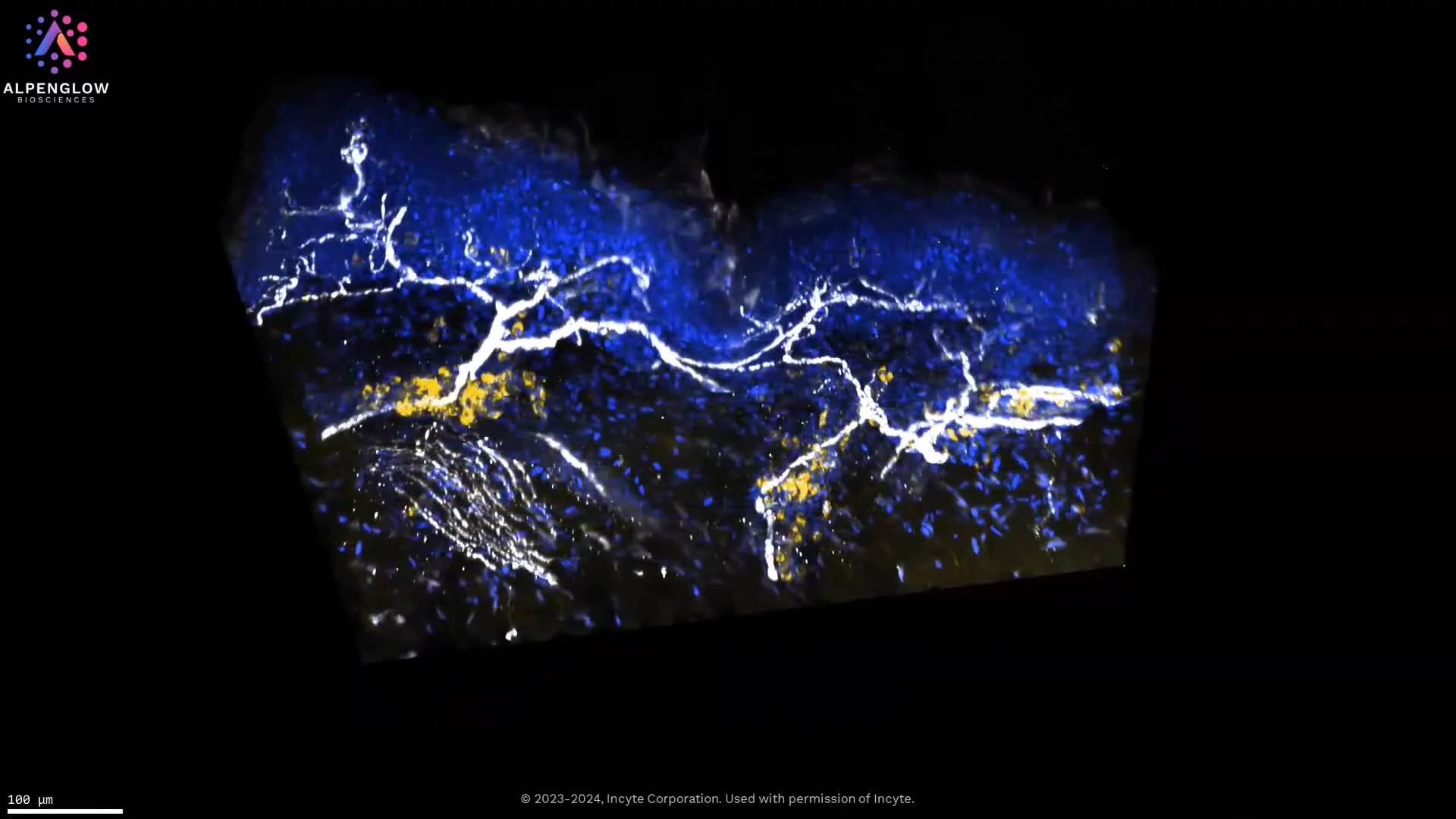
3D imaging
Intact architecture across tissue depth
Volumetric imaging keeps structures connected, helping researchers evaluate biology in the context where it occurs.
Quantitative analysis
Spatial data ready for deeper interpretation
AI-powered analysis can turn 3D datasets into measurable features for spatial biology and digital pathology workflows.
The 2D limitation
Thin sections can simplify biology that is fundamentally three-dimensional.
A 2D section can be powerful for cell and tissue review, but it captures a narrow plane through a larger specimen. When the biology depends on depth, proximity, branching, continuity, or rare features, spatial context can be lost.
Small sampling window
A thin section may not represent the full tissue volume, especially when structures are sparse, unevenly distributed, or localized in specific regions.
Depth is collapsed
Cells or structures that appear close in a plane may be separated across depth, while important signals above or below the section may be missed.
Continuity is broken
Networks such as vessels, nerves, follicles, glands, and immune structures are easier to interpret when their connected 3D architecture is preserved.
3D tissue imaging helps preserve the relationships that 2D sections can fragment, creating a stronger foundation for spatial profiling, digital pathology, and quantitative tissue analysis.
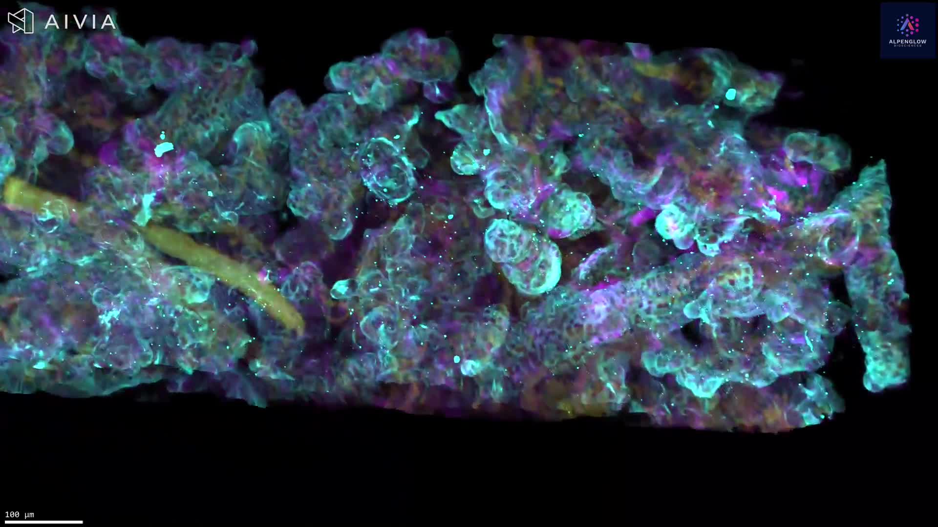
From plane to volumeWhat 3D preserves
3D tissue imaging keeps biology connected, measurable, and spatially interpretable.
By imaging intact samples as volumetric datasets, researchers can study tissue architecture across depth instead of relying on isolated planes. This creates a stronger foundation for spatial profiling, digital pathology, AI-powered analysis, and quantitative tissue analysis.
From isolated sections to whole tissue context.
3D imaging helps preserve the spatial relationships between cells, vessels, nerves, glands, immune structures, and tissue compartments. That context can be critical when morphology, proximity, branching, or depth drives the biological question.
The shift is not just visual. Whole tissue imaging can support more complete interpretation of where structures are, how they connect, and how they relate across the sample.
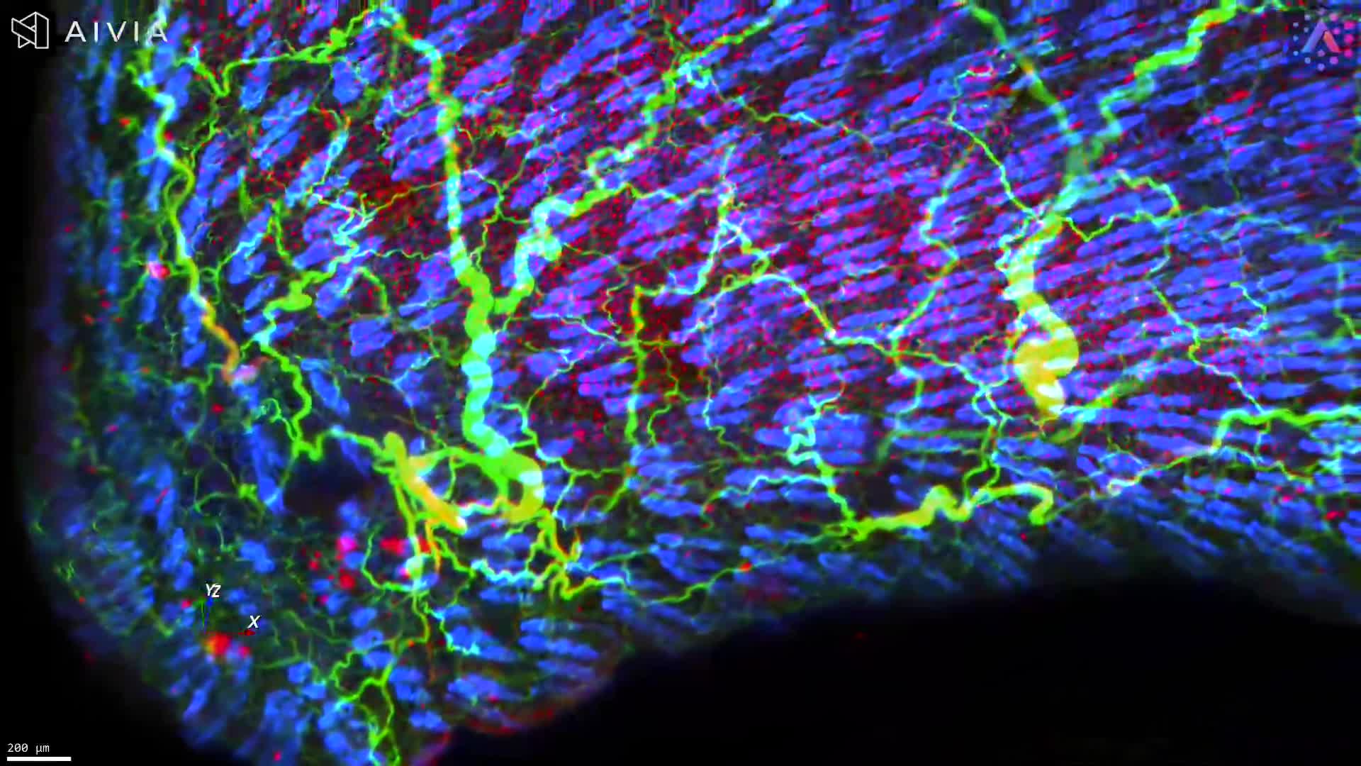
Architecture
Evaluate tissue structures as connected systems rather than disconnected cross sections.
Depth
View biology above, below, and between planes to understand what a single section may miss.
Proximity
Measure spatial relationships between cells, structures, and compartments in 3D context.
Continuity
Follow branching and connected features such as vessels, nerves, follicles, glands, and immune structures.
The result is richer spatial information from intact tissue. 3D tissue imaging can support both qualitative review and quantitative analysis of complex biology.
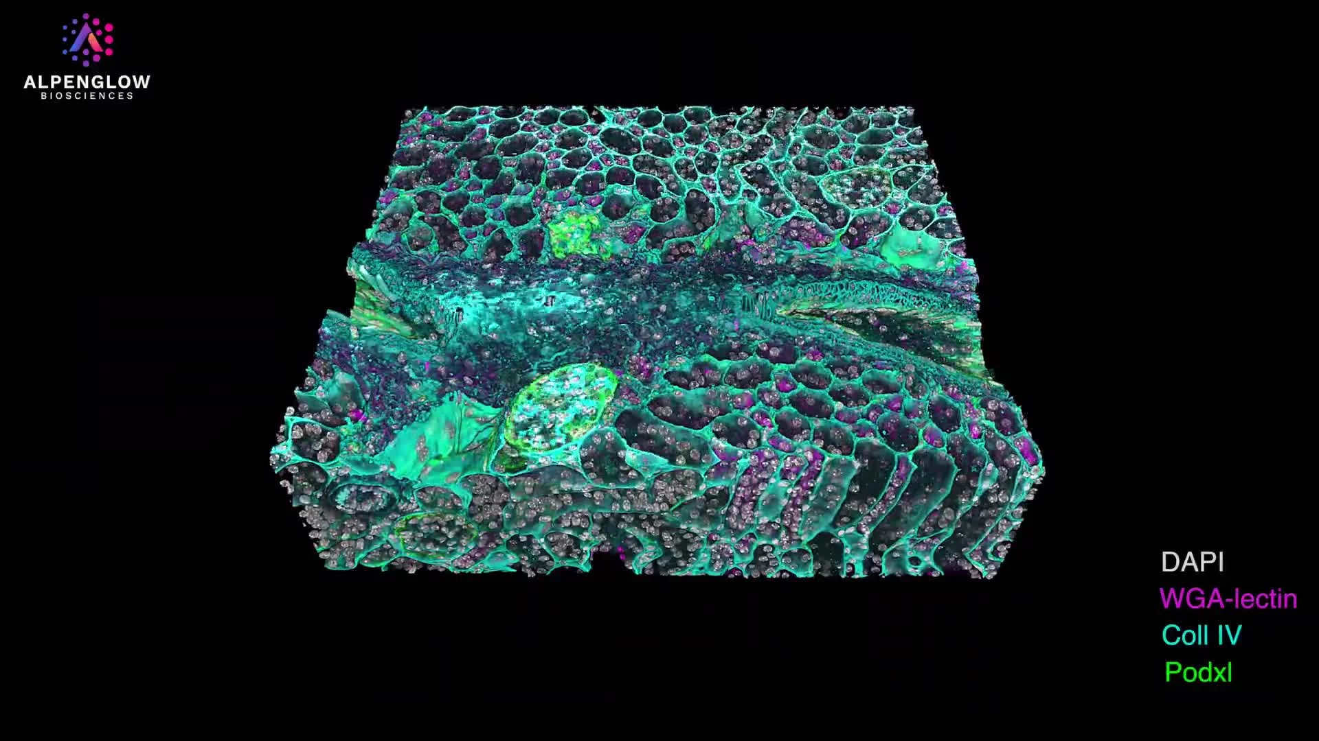
Spatial context preservedKey biological applications
Where 3D tissue imaging becomes essential to interpretation.
Some biological questions depend on structures and spatial relationships that are difficult to evaluate from isolated sections alone. 3D tissue imaging supports whole tissue imaging, spatial profiling, volumetric datasets, digital pathology, AI-powered analysis, and quantitative tissue analysis from intact samples.
Convoluted structures
Vessels, nerves, glands, follicles, and crypts often require 3D context to evaluate shape, branching, and continuity.
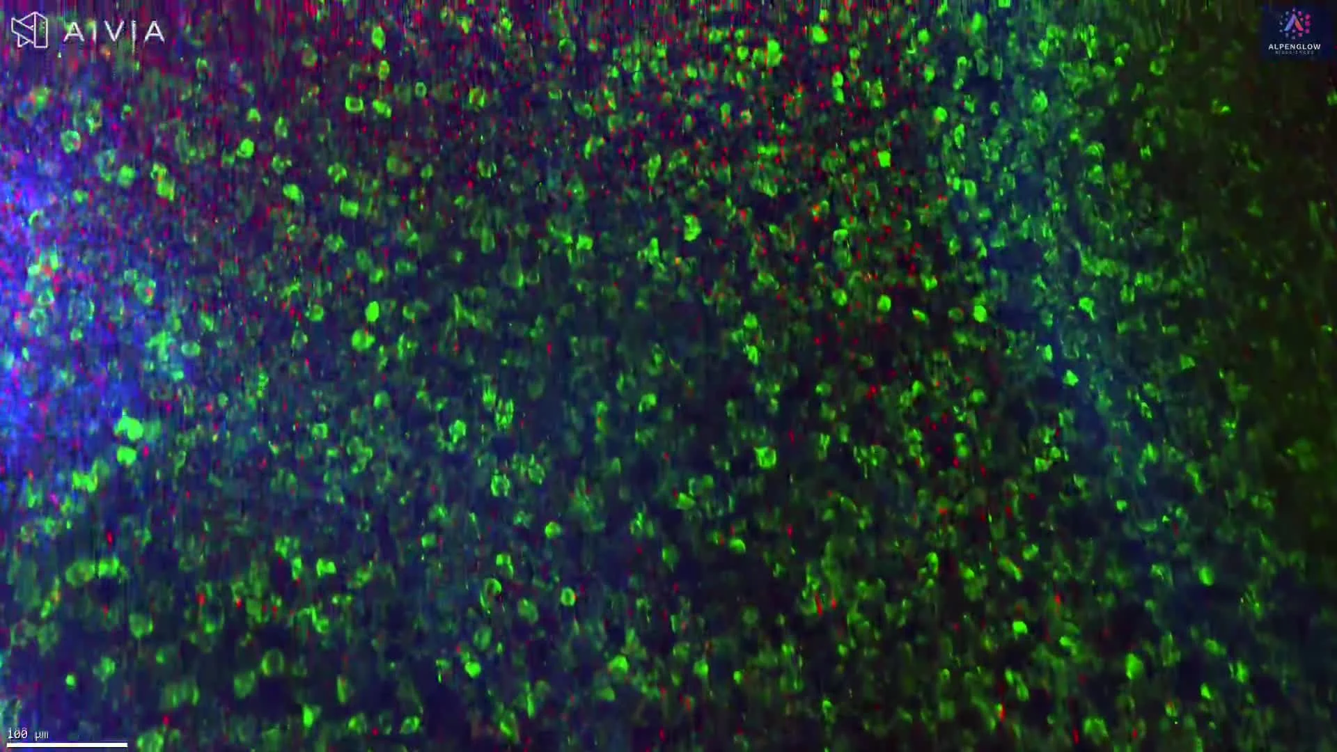
Complex cell distributions
Immune cells, tumor regions, and stromal compartments can be interpreted in relation to nearby structures across tissue depth.
Sparse biological features
Rare cells, focal lesions, tertiary lymphoid structures, and localized features can be missed when sampling is limited.
Tissue-scale architecture
Whole tissue imaging helps reveal how compartments, regions, and structures are organized across the sample.
Who benefits?
Built for teams that need more than a representative section.
Translational research teams, drug discovery groups, spatial biology researchers, and digital pathology innovators use 3D tissue imaging to study intact tissue context, generate volumetric datasets, and support AI-powered analysis for quantitative tissue analysis.

From tissue to data
How does Alpenglow turn intact tissue into quantitative 3D data?
Alpenglow’s Aurora 3D™ Spatial Biology Solution connects 3D tissue imaging, volumetric data processing, and AI-powered analysis so researchers can generate spatial profiling data from intact tissue samples.
Answer in brief
Image the tissue, manage the volume, quantify the biology.
The workflow is designed to move from whole tissue imaging to analysis-ready volumetric datasets, preserving spatial context for digital pathology, spatial biology, and quantitative tissue analysis.
3Di™ images intact tissue
Light-sheet microscopy captures tissue architecture in 3D.
3Dm™ manages the data
Large volumetric datasets are prepared for review and analysis.
3Dai™ quantifies features
AI-powered analysis supports measurement of cells, structures, and spatial relationships.
What is the Aurora 3D™ Spatial Biology Solution?
Aurora is Alpenglow’s end-to-end platform for 3D tissue imaging, data management, and AI-powered analysis. It is designed to help researchers study intact tissue context and generate quantitative tissue analysis from volumetric datasets.
How does 3Di™ HOTLS microscopy support whole tissue imaging?
3Di™ HOTLS microscopy uses light-sheet imaging to capture intact samples in 3D, helping preserve tissue-scale architecture, depth, continuity, and spatial relationships that can be difficult to evaluate from thin sections alone.
Why are 3Dm™ and 3Dai™ important after imaging?
3Dm™ supports management and processing of large 3D datasets, while 3Dai™ applies AI-powered analysis to quantify cells, structures, tissue features, and spatial relationships for digital pathology and spatial profiling workflows.
What can researchers measure with 3D tissue imaging?
Researchers can use 3D tissue imaging to study cell distributions, vessel and nerve networks, glandular and follicular structures, immune organization, tissue compartments, rare features, and quantitative spatial relationships across intact tissue.
Explore the Alpenglow workflow for 3D spatial biology.
Learn how Aurora 3D™ connects 3D tissue imaging, spatial profiling, digital pathology, volumetric datasets, and AI-powered quantitative tissue analysis.
Move from sections to spatial insight
Ready to see what 3D tissue imaging can reveal in your samples?
Explore how Alpenglow supports whole tissue imaging, spatial profiling, volumetric datasets, digital pathology, AI-powered analysis, and quantitative tissue analysis with the Aurora 3D™ Spatial Biology Solution.