
3D tissue imaging microscope
3Di™ Hybrid Open-Top Light-Sheet Microscope
A dedicated instrument for high-resolution 3D tissue imaging, whole tissue imaging, and quantitative tissue analysis across intact biological specimens.
What the microscope does
The 3Di™ system converts intact biological specimens into quantitative 3D datasets.
The 3Di™ microscope is built for high-resolution 3D tissue imaging, whole tissue imaging, and spatial biology workflows. It helps researchers move from large-volume sample context to detailed regions of interest while preserving the architecture needed for digital pathology, spatial profiling, and quantitative tissue analysis.
Image large tissue volumes
Acquire complete 3D images of intact biological tissues while maintaining whole sample context across large volumetric datasets.
Identify regions of interest
Use overview imaging to navigate the sample, locate meaningful tissue structures, and select regions for deeper high-resolution analysis.
Capture submicrometer detail
Move from broad tissue architecture to fine structural detail, supporting AI-powered analysis and quantitative tissue readouts.
Scout-to-Zoom workflow
Whole tissue context first. High-resolution 3D detail where the biology matters.
The 3Di™ microscope combines rapid Scout imaging with high-resolution Zoom imaging, helping researchers move from large intact tissue volumes to selected regions of interest within a single 3D tissue imaging workflow.
Scout the whole sample
Capture a rapid overview of the intact specimen to preserve whole tissue imaging context across large volumetric datasets.
Select regions of interest
Navigate the tissue in 3D, locate relevant structures, and choose regions for deeper spatial profiling and digital pathology analysis.
Zoom into submicrometer detail
Acquire high-resolution 3D images of selected regions while maintaining the larger biological context from the Scout scan.
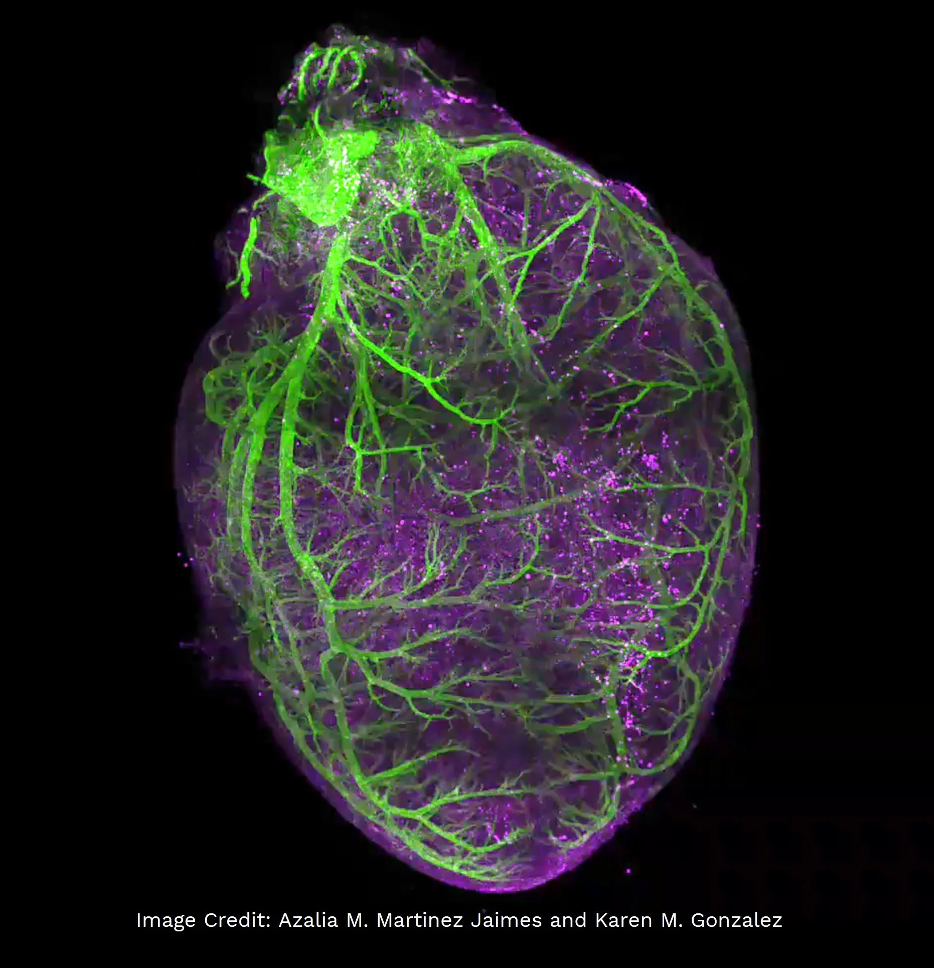
Whole rat heart overview
Rapid low-resolution imaging provides whole-sample context and navigation.
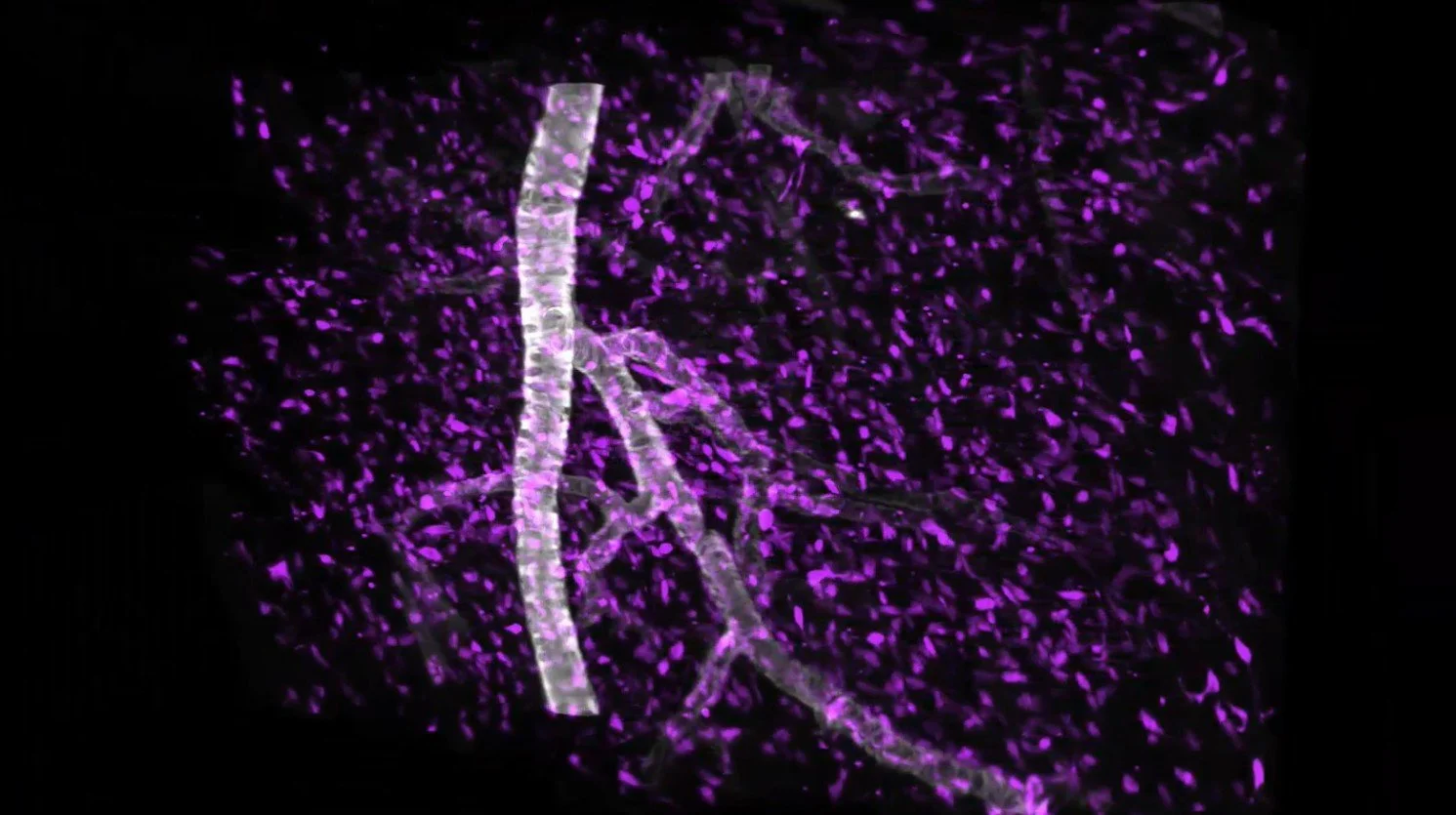
ROI from the whole rat heart
High-resolution imaging captures local structural detail inside selected tissue regions.
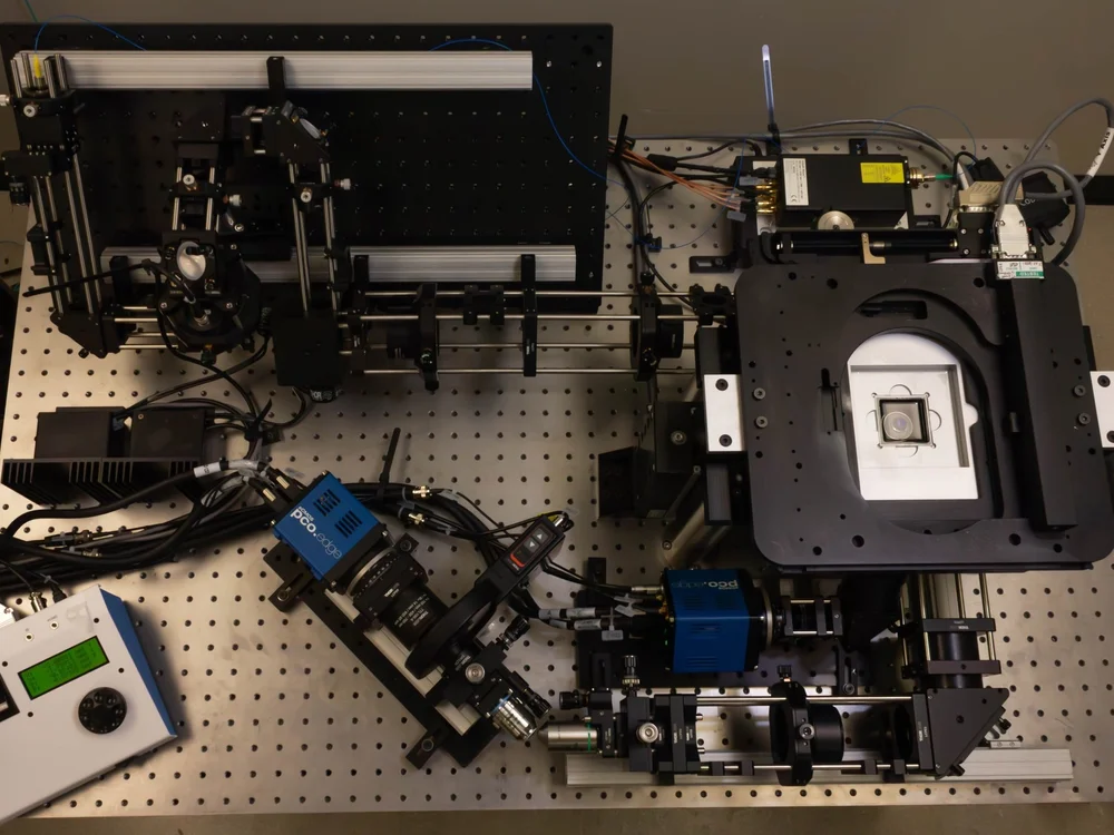
HOTLS architecture
Peer-reviewed optical architecture for multi-scale cleared tissue imaging.
The 3Di™ microscope is built on a Hybrid Open-Top Light-Sheet architecture described in Nature Methods. The design combines dedicated illumination and collection optics to support versatile multi-scale imaging of cleared tissues.
Publication reference
A hybrid open-top light-sheet microscope for versatile multi-scale imaging of cleared tissues
Nature Methods, May 2022. Peer-reviewed technical foundation for the HOTLS architecture.
Read publication →
Hybrid Open-Top Light-Sheet optical layout
Microscope schematic showing the optical architecture behind the published HOTLS design.
Open-top sample flexibility
Flexible access for diverse intact tissue imaging workflows.
The 3Di™ microscope’s open-top design provides practical access to samples and supports multiple holder configurations for a wide range of specimen shapes, sizes, and preparation methods.
Open-top access
Practical sample loading and positioning for intact tissue imaging workflows.
Flexible access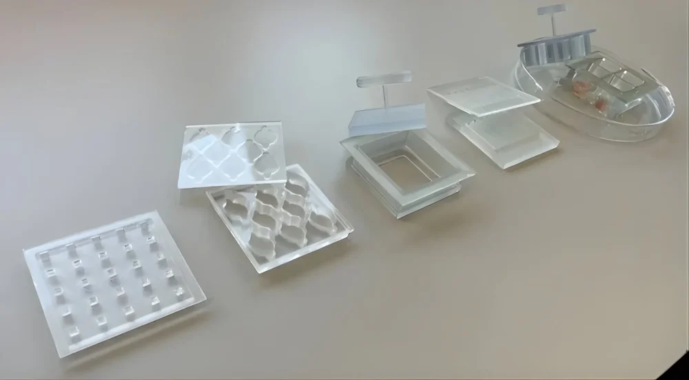
Holder configurations
Holder options support diverse specimen formats and experimental needs.
Sample formatsEasy sample access
The open-top format supports straightforward sample loading, positioning, and handling for cleared tissue imaging.
Flexible holder configurations
Sample holders support different specimen formats, including organoids, biopsies, tissue blocks, and whole organs.
Multiple specimen workflows
Multi-specimen scanning supports more efficient acquisition when imaging multiple samples in the same session.
Large scan area
The microscope’s large scan area, up to 12 cm × 7.5 cm × 1 cm, enables the acquisition of complete 3D images of large biological tissues.
LUMI interface
Intuitive control for complex 3D imaging workflows.
LUMI simplifies scan setup, live preview, and real-time visualization for the 3Di™ microscope, helping researchers operate Scout and Zoom imaging modes within a streamlined interface.
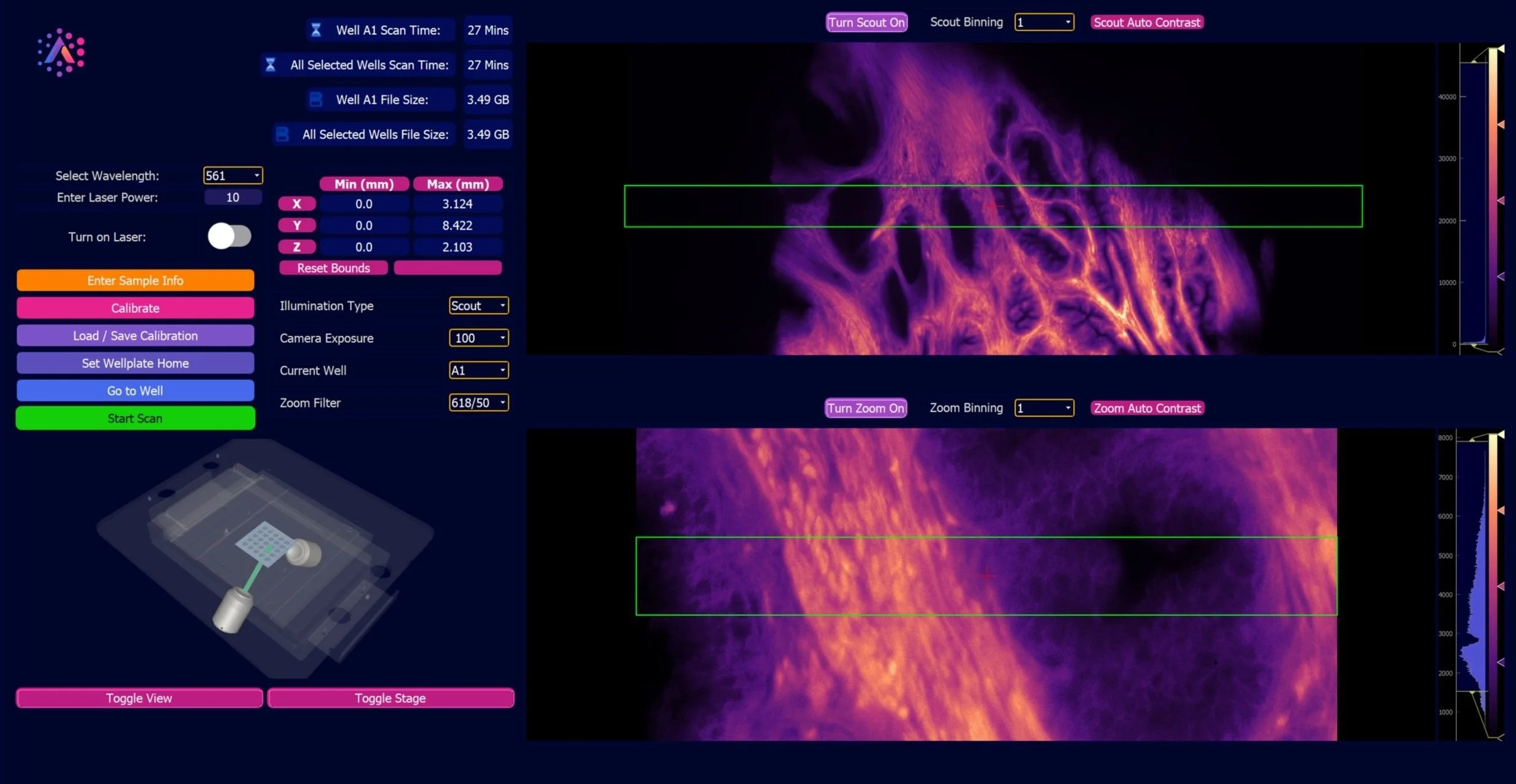
Scan setup, preview, and acquisition in one interface
Live visualization supports both Scout and Zoom imaging workflows.
Intuitive scan setup
The interface streamlines scan setup so users can configure imaging workflows with fewer manual steps.
Live preview
Simultaneous live previews in Scout and Zoom modes support real-time visualization before acquisition.
Easy interface
A user-friendly interface helps researchers navigate and control the system across 3D tissue imaging workflows.
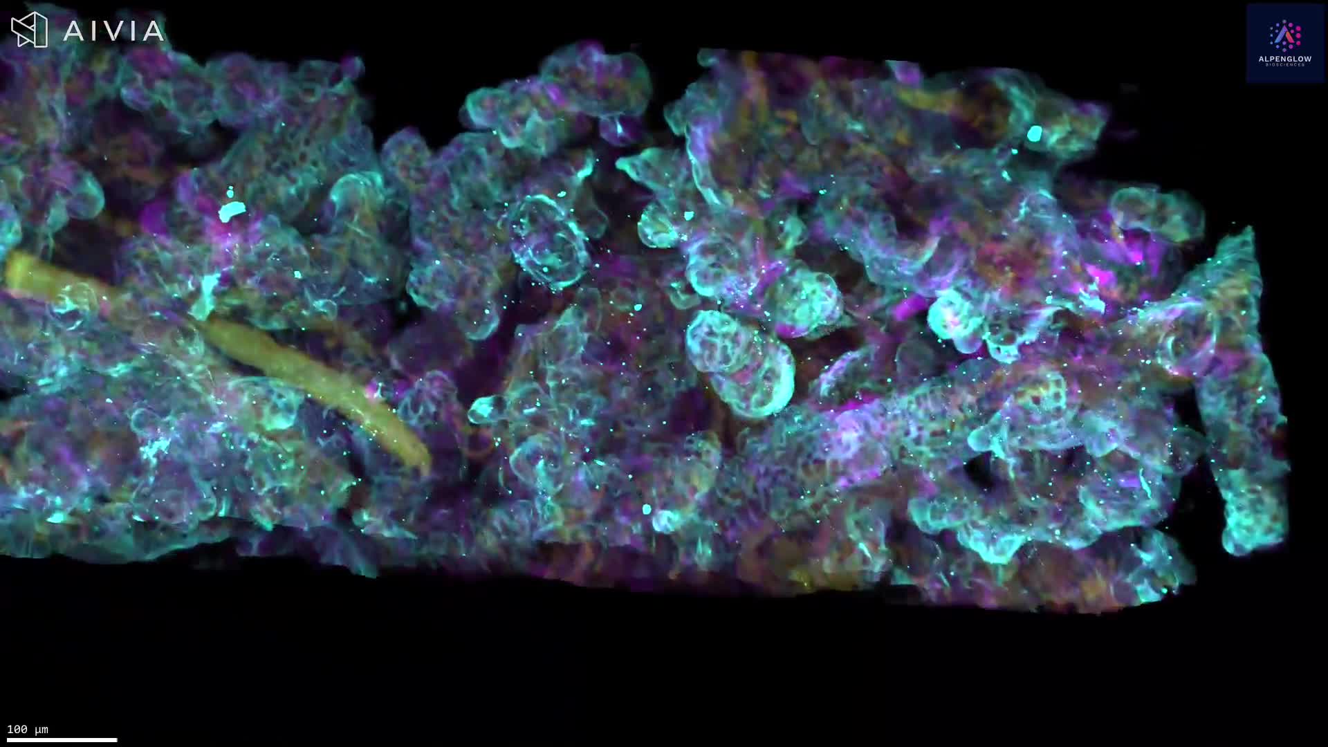
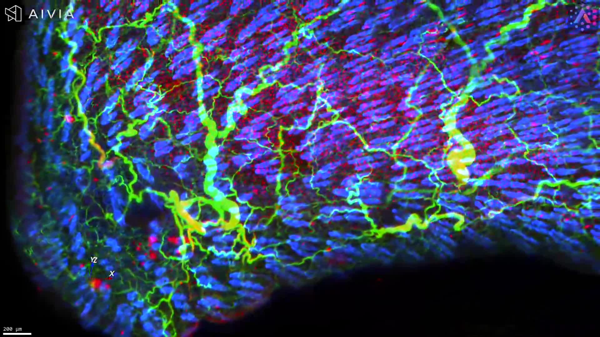
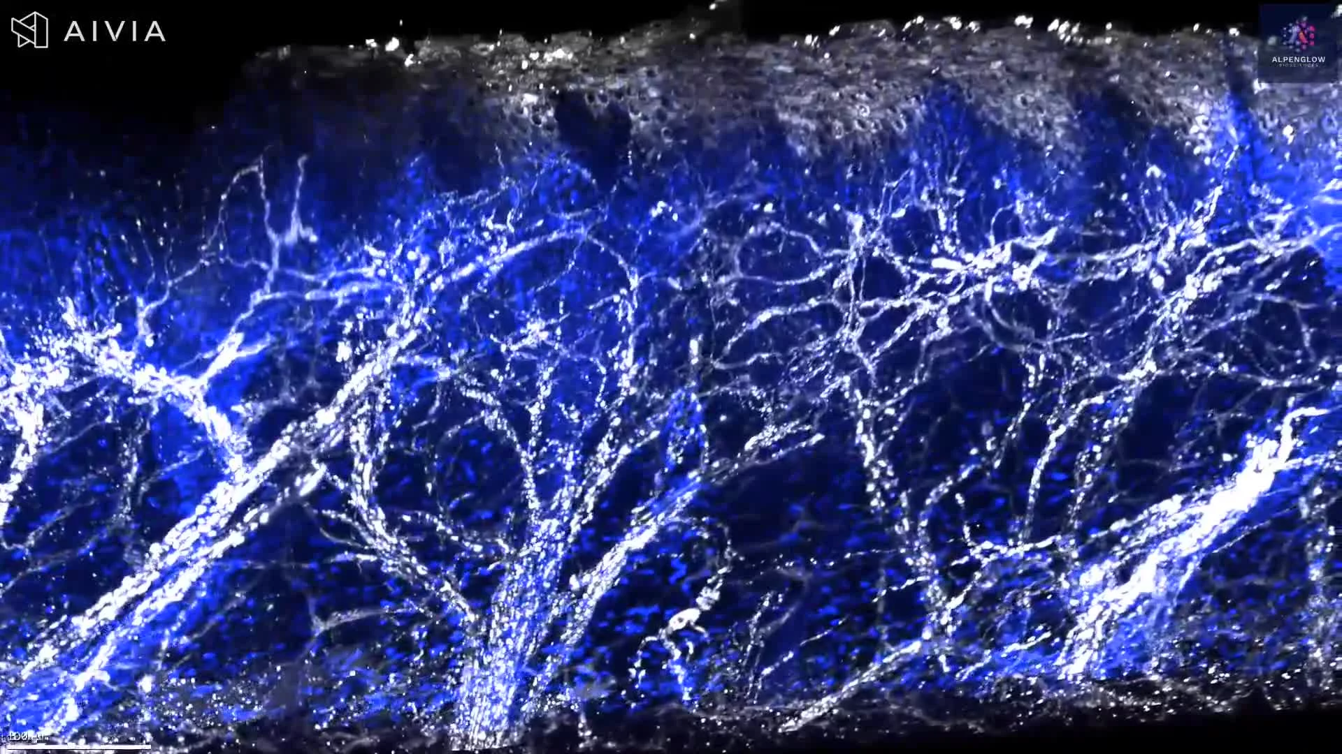
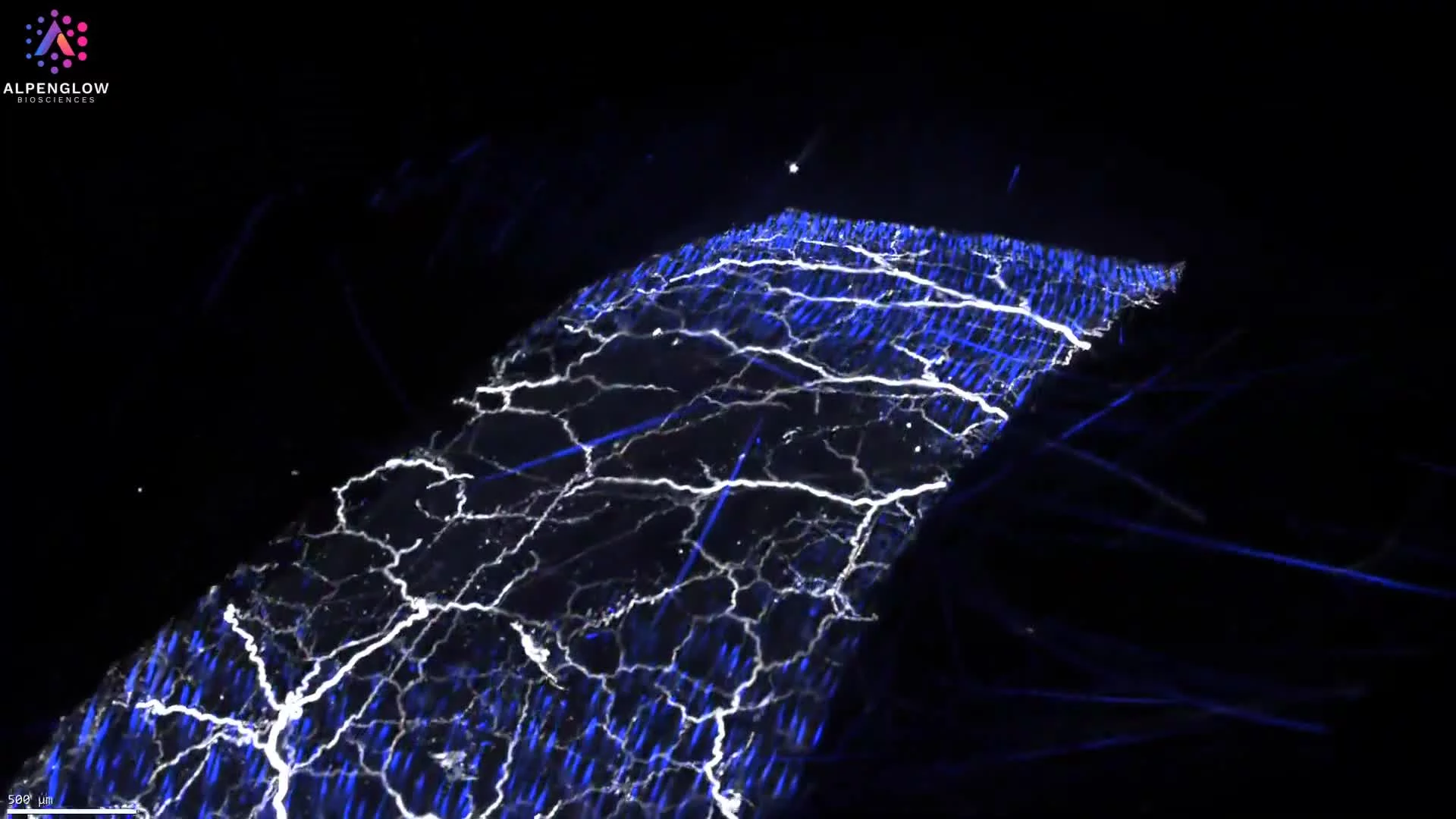
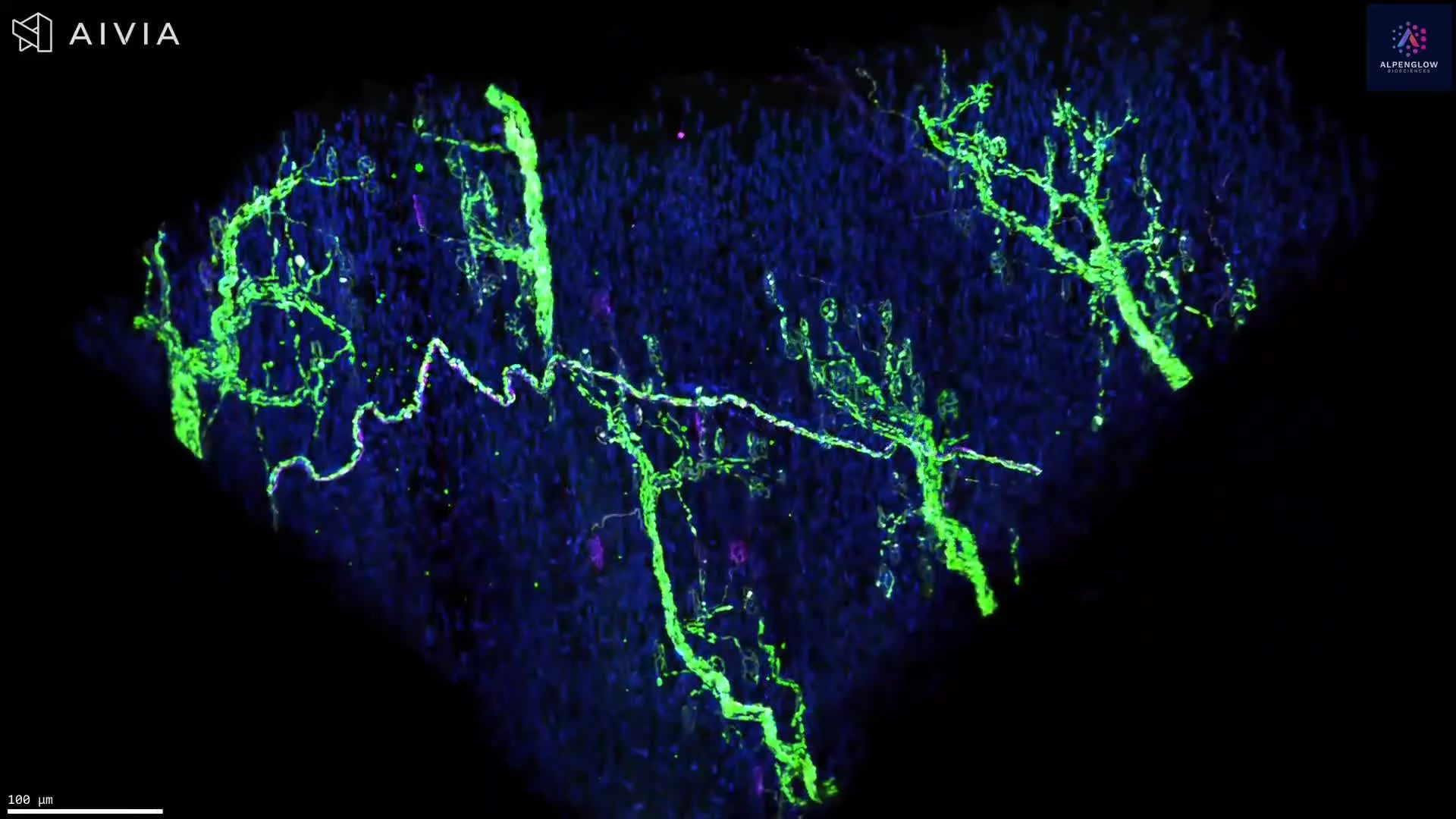
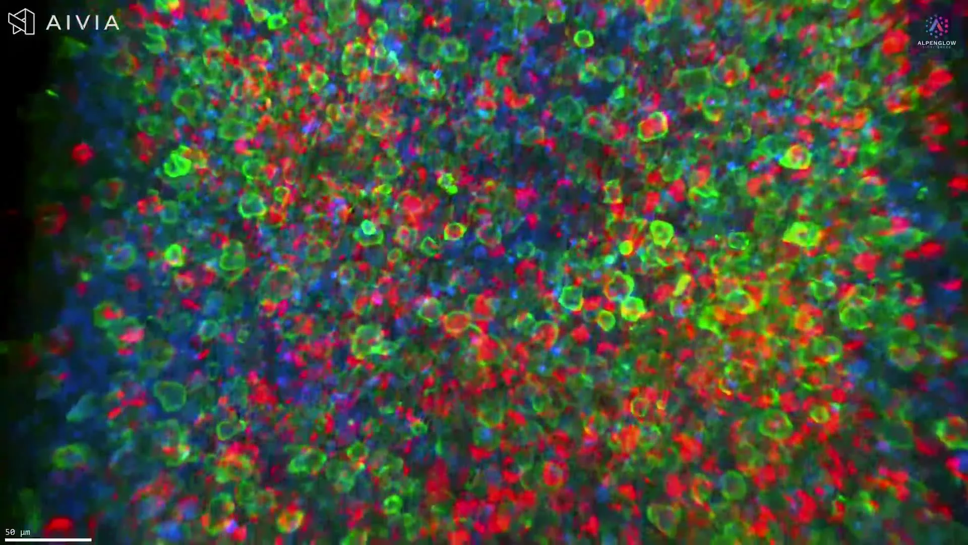
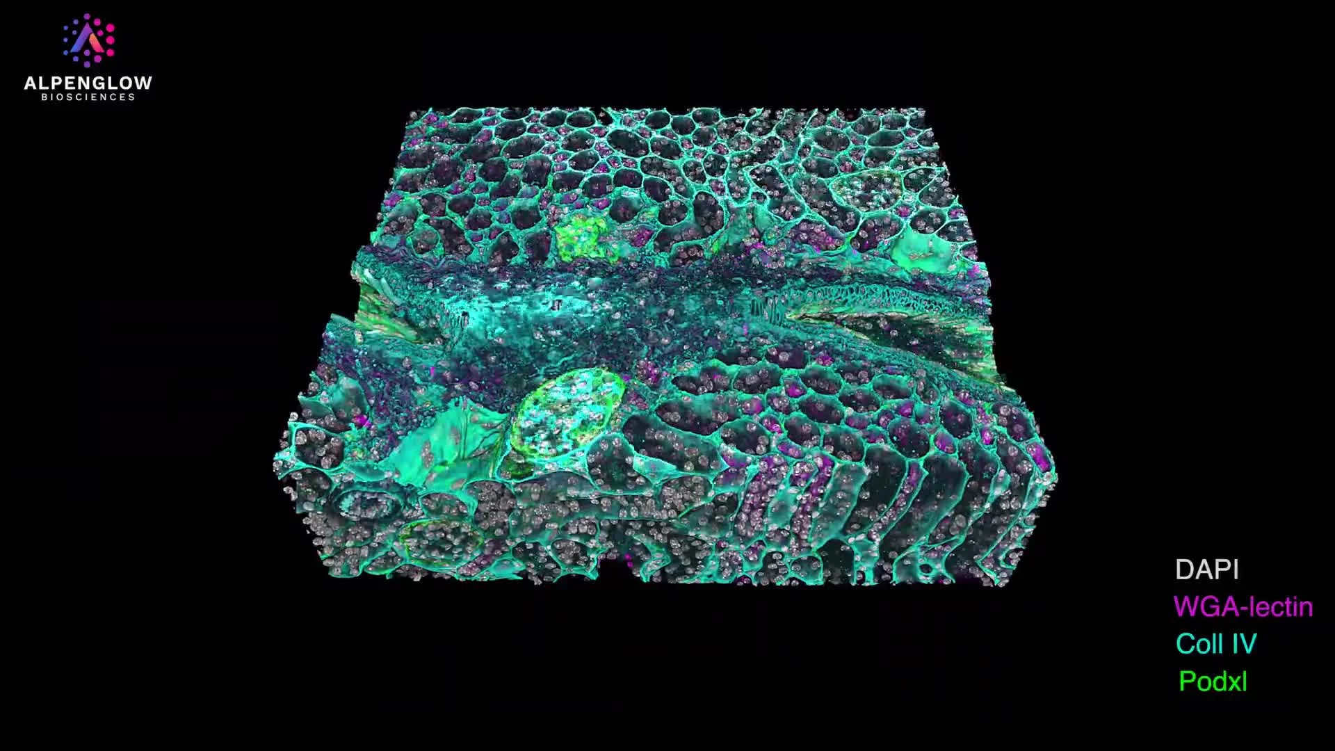
Generated datasets
See the volumetric datasets produced on the 3Di™ HOTLS microscope.
Explore examples of 3D tissue imaging across intact biological specimens, from large-volume context to high-resolution structural detail.
Technical specifications
Built for large volume, high-resolution 3D tissue imaging.
The 3Di™ HOTLS microscope combines a large scan area, Scout-to-Zoom imaging, multi-channel fluorescence, and LUMI control software for intact tissue imaging workflows.
3Di™ technical specifications
HOTLS microscope| Specification | Value |
|---|---|
| Format | Hybrid Open-Top Light-Sheet |
| Maximum specimen size | Up to 12 cm × 7.5 cm × 1 cm (x, y, z) |
| Maximum collection | 0.7 NA |
| Maximum illumination | 0.06 NA |
| Scout resolution | 2 µm/pixel |
| Zoom resolution | 0.17 µm/pixel |
| Low power resolution magnification | 2.5 X |
| High power resolution magnification | 40 X |
| Laser lines standard, now up to 5 channels | 405 nm, 488 nm, 561 nm, 638 nm (735 nm, 780 nm) |
| Camera | 2 × Hamamatsu ORCA Fusion BT |
| Refractive index capability | RI 1.33 to 1.56 |
| Multi well, multi-specimen scanning | Yes |
| Microscope control software | LUMI Graphical User Interface |