
Immuno-oncology applications
3D immuno-oncology imaging for full-volume tumor immune analysis
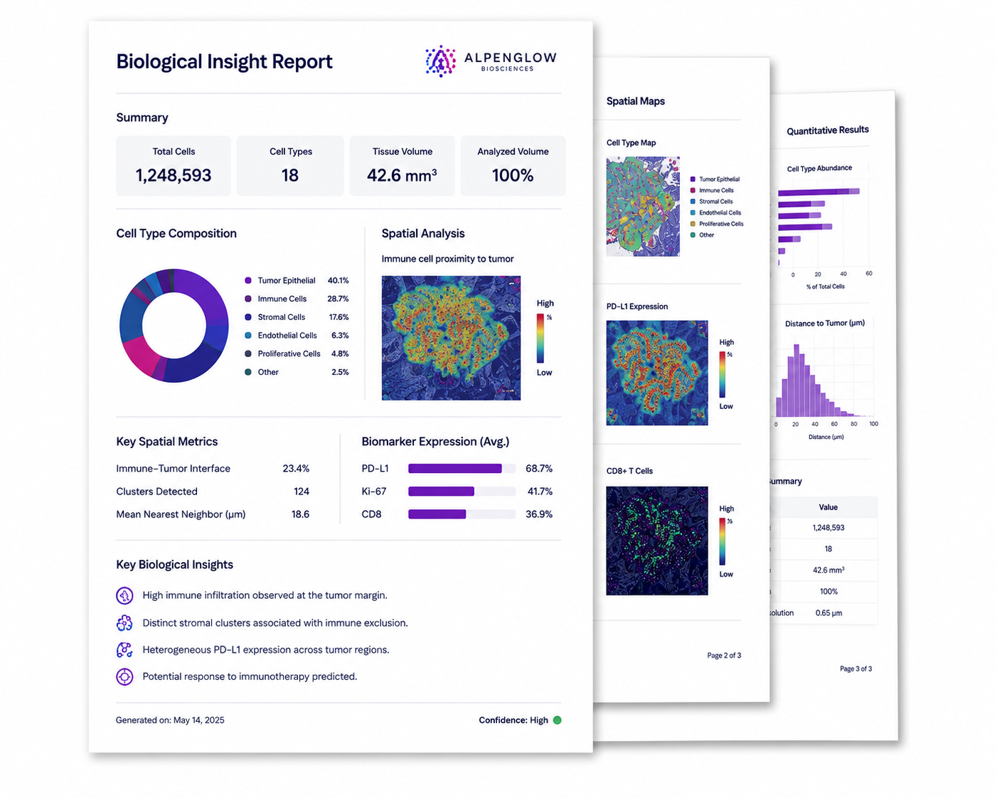
The 3D I/O Pro™ Assay combines high-resolution 3D tissue imaging, digital pathology, and AI-powered analysis to map immune exclusion, lymphocyte distribution, tertiary lymphoid structures, collagen organization, and tumor microenvironment architecture across intact tumor samples.
3D I/O Pro™ Assay
A 3D digital pathology assay for the tumor immune microenvironment
The 3D I/O Pro™ Assay combines high-resolution 3D tissue imaging with quantitative tissue analysis to measure immune contexture, lymphocyte organization, immune exclusion, collagen structure, and tertiary lymphoid structures across intact tumor samples.
Why 3D tumor immune microenvironment imaging matters
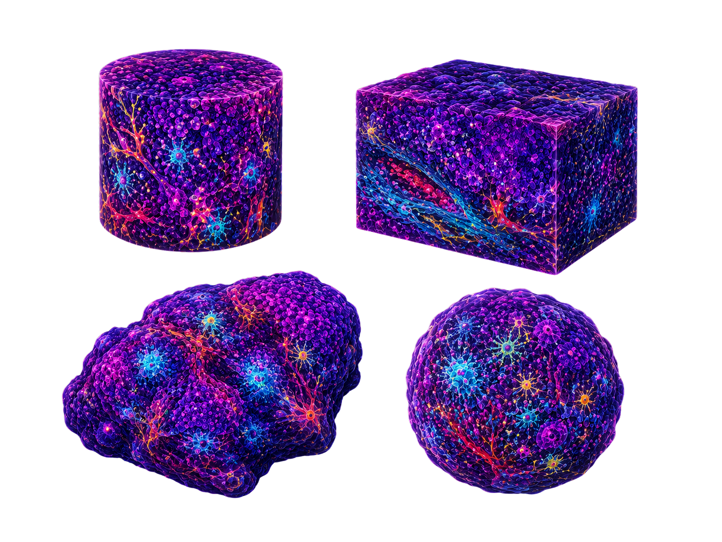
Tumor immunity is spatial. Thin sections can miss the pattern.
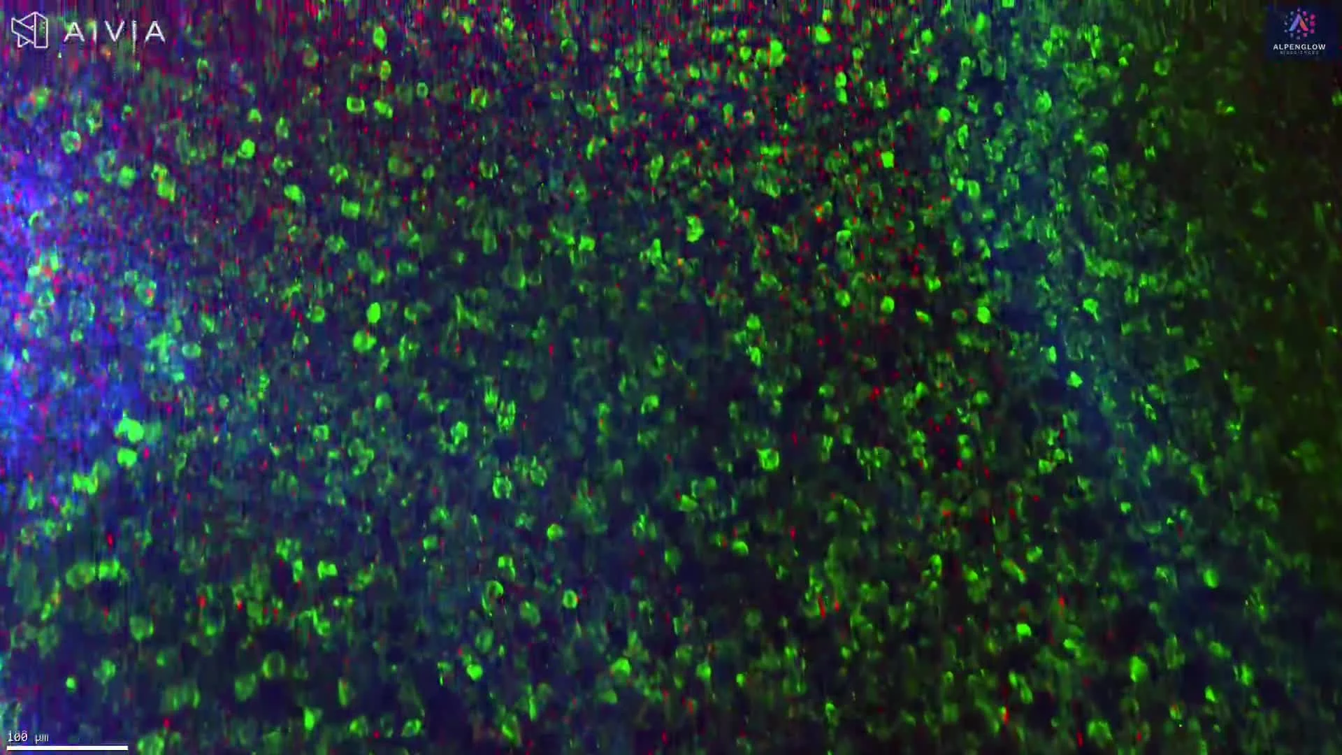
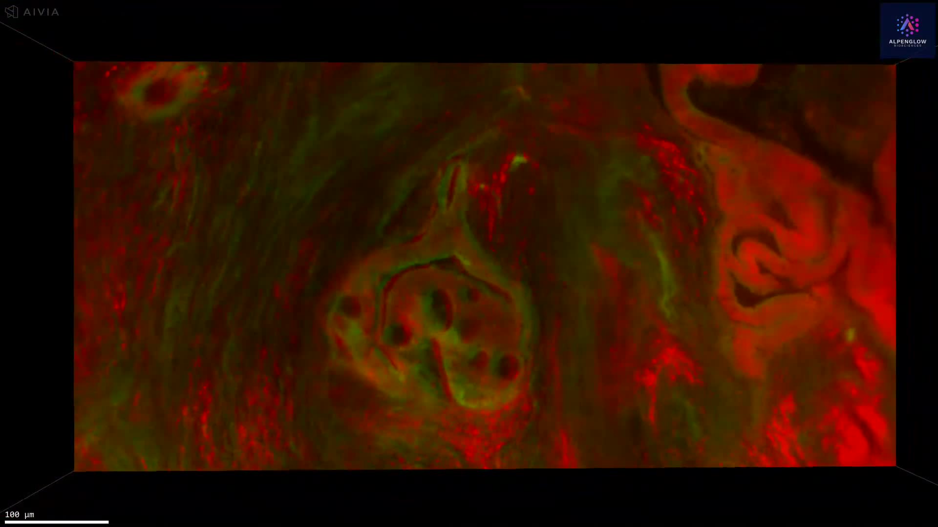
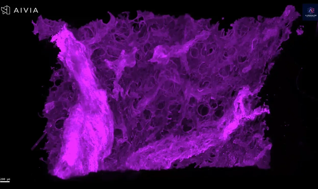
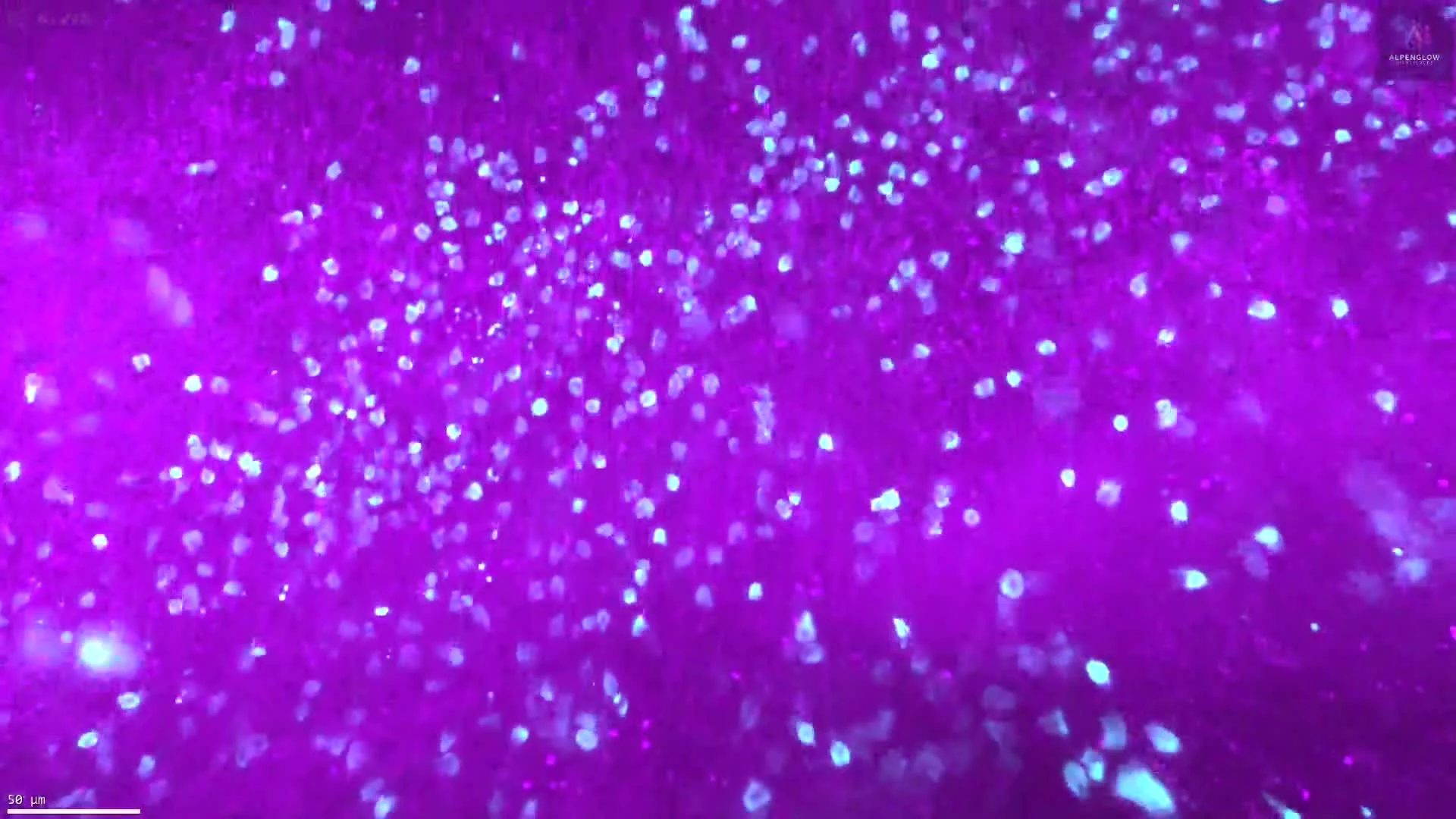
Solid tumors contain lymphocyte gradients, immune-excluded regions, collagen-rich stroma, tertiary lymphoid structures, and local tumor-immune interactions. 3D tissue imaging captures these relationships across the tumor volume.
Traditional workflows
Limited views of a heterogeneous tumor
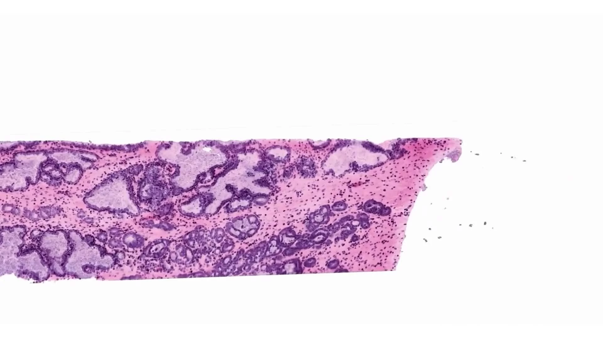
Single 2D sections can provide useful histology, but they may sample a small part of the tumor and separate features that are connected in 3D.
- Small fraction of the tumor volume
- Single-plane view of lymphocyte distribution
- TLS, collagen structure, and immune exclusion can be harder to interpret in context
3D tissue imaging
Full-volume insight for quantitative immuno-oncology
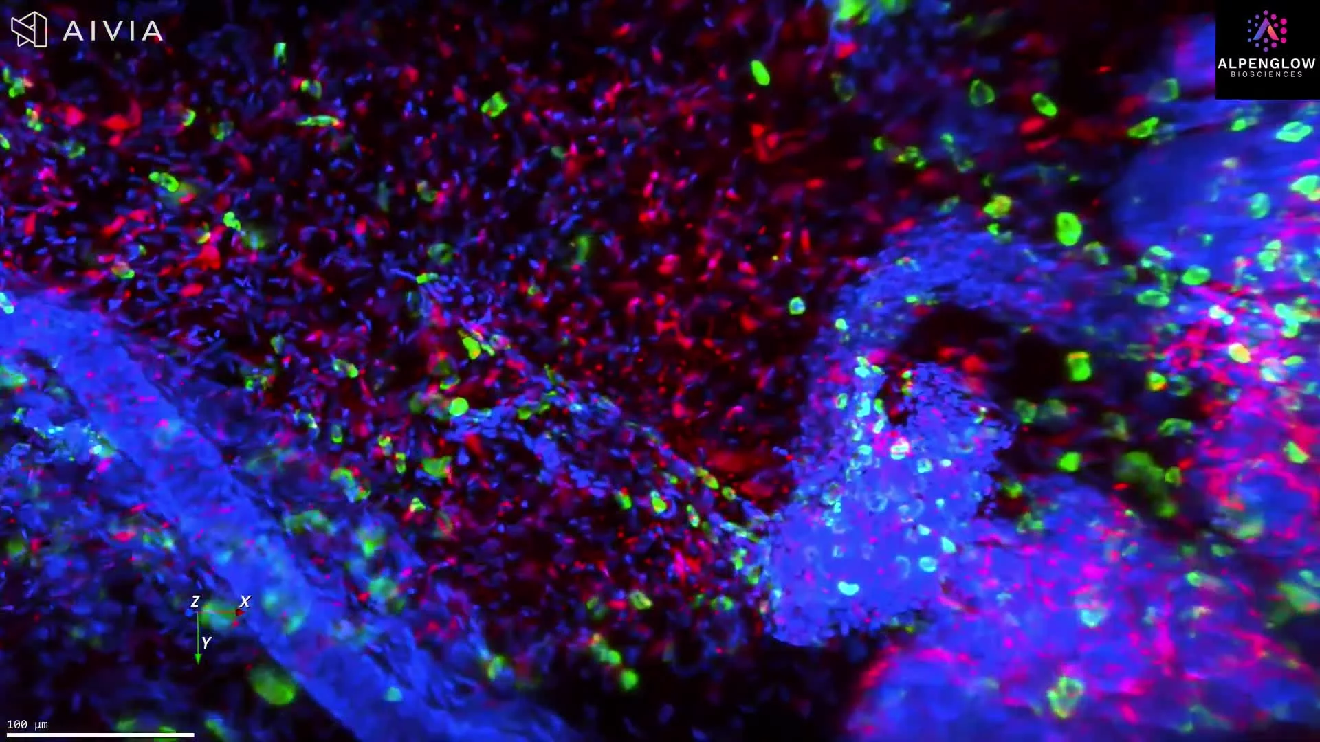
Whole tissue imaging preserves spatial context, enabling quantitative tissue analysis of immune organization, tumor architecture, stromal structure, and cell relationships across intact tumor samples.
- Lymphocyte gradients from tumor periphery to core
- Immune exclusion across tumor margin, core, and stroma
- TLS volume, shape, location, and proximity to tumor nests
From qualitative impressions to reproducible metrics. The 3D I/O Pro™ Assay combines whole tissue imaging with quantitative analysis to support mechanism-of-action studies, spatial biomarker discovery, and translational immuno-oncology research.
3D I/O Pro™ workflow
Immuno-oncology assays built on the Aurora 3D™ workflow
Start with intact tumor tissue, capture high-resolution 3D tissue imaging, process large volumetric datasets, and apply AI-powered analysis for quantitative immuno-oncology readouts.
Input
Intact tumor tissue
Tumor samples and biopsies prepared for whole tissue 3D imaging.
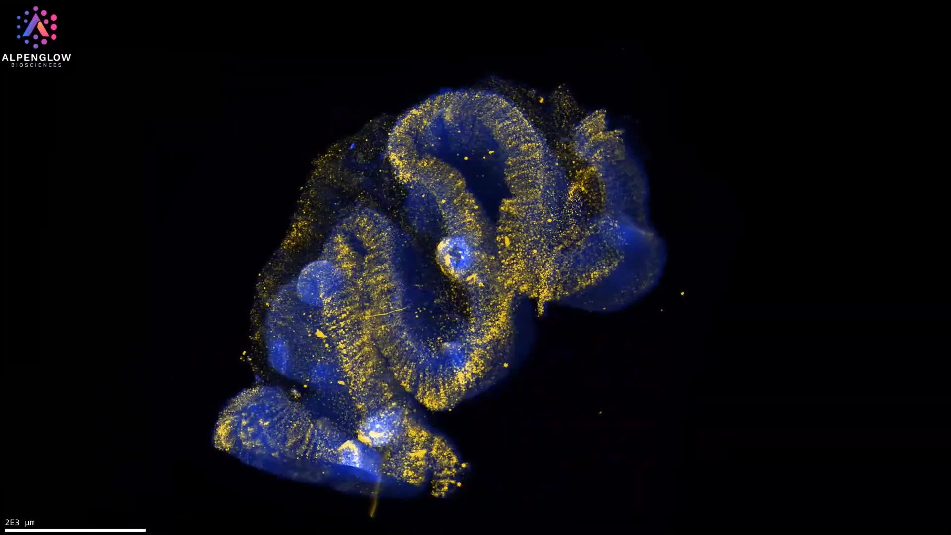
3D imaging
3Di HOTLS
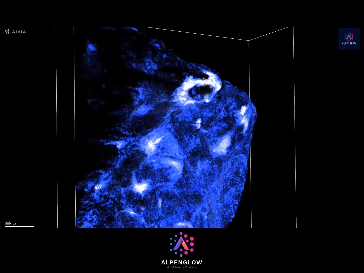
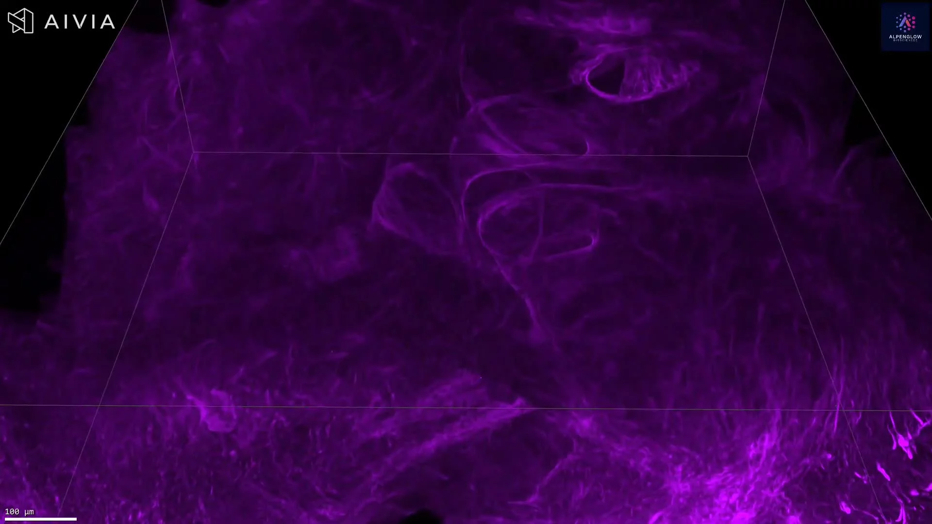
High-resolution 3D tissue imaging across intact immuno-oncology samples.
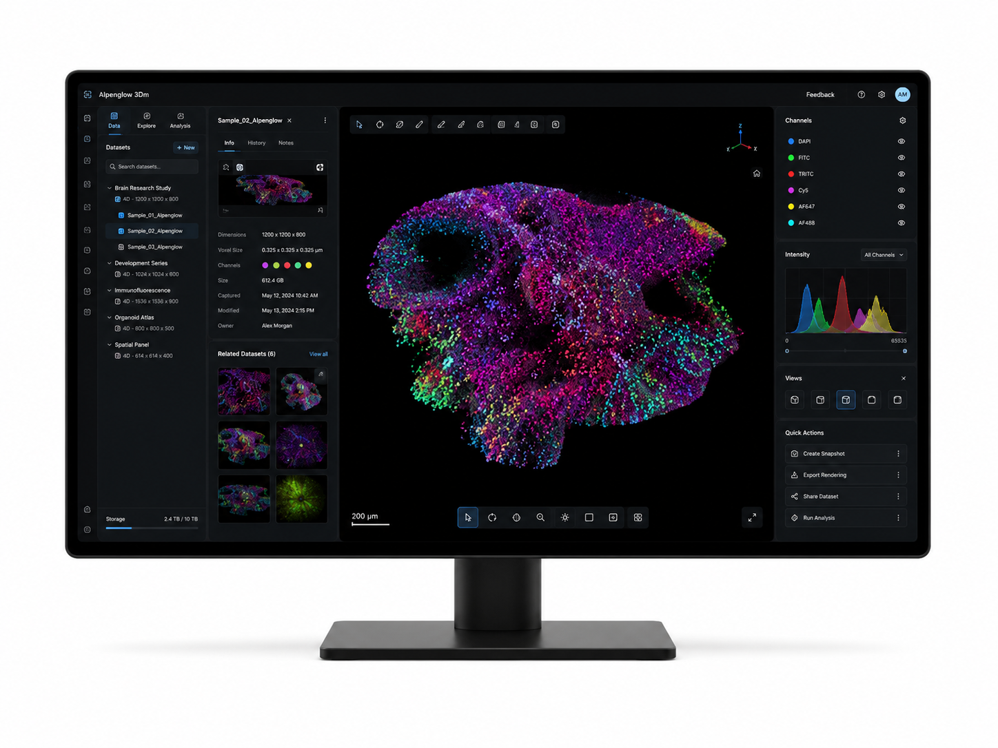
Data management
3Dm
Processing, correction, organization, and preparation of large 3D datasets.
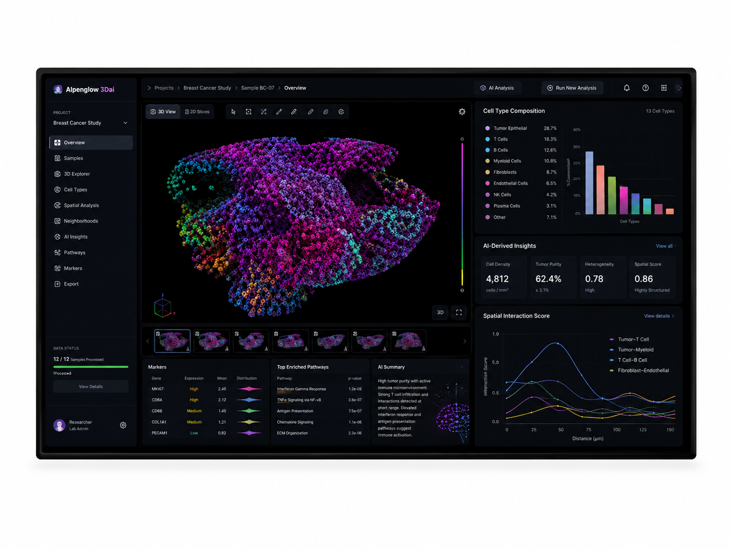
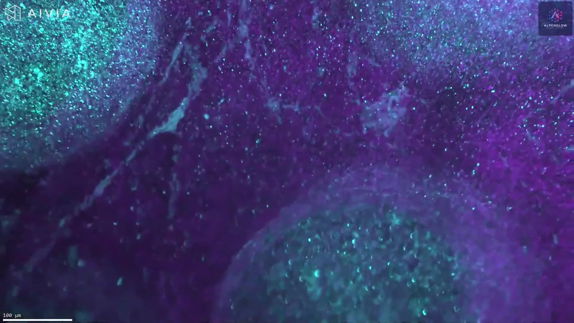
AI analysis
3Dai
AI-powered analysis for tumor regions, immune cells, TLS, collagen, and quantitative tissue analysis.
Output
IO insights
Quantitative readouts for immune exclusion, lymphocyte spatial mapping, TLS organization, collagen structure, and tumor immune contexture.
Marker panels
Flexible panels for tumor immune microenvironment profiling
Different immuno-oncology programs require different panels. The 3D I/O Pro™ Assay supports pre-optimized and custom marker strategies for 3D tissue imaging, spatial profiling, and quantitative tissue analysis.
Option A
Select a pre-optimized panel
Use an immuno-oncology-focused marker panel to support tumor identification, lymphocyte mapping, and spatial analysis across intact tumor samples.
Example markers shown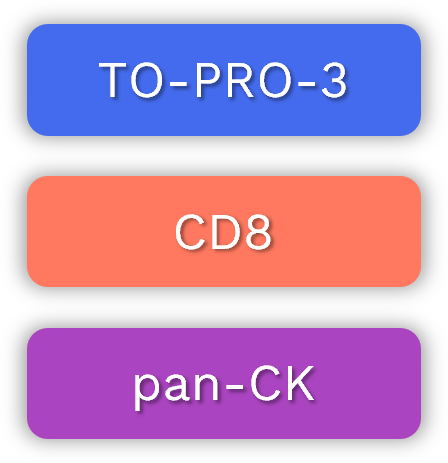
Option B
Design your panel
Adapt the marker strategy to your tumor type, species, experimental goals, and immuno-oncology endpoint, including immune exclusion, TLS, collagen, and stromal structure.
Custom marker strategy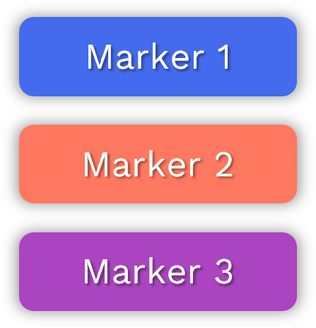
Example datasets
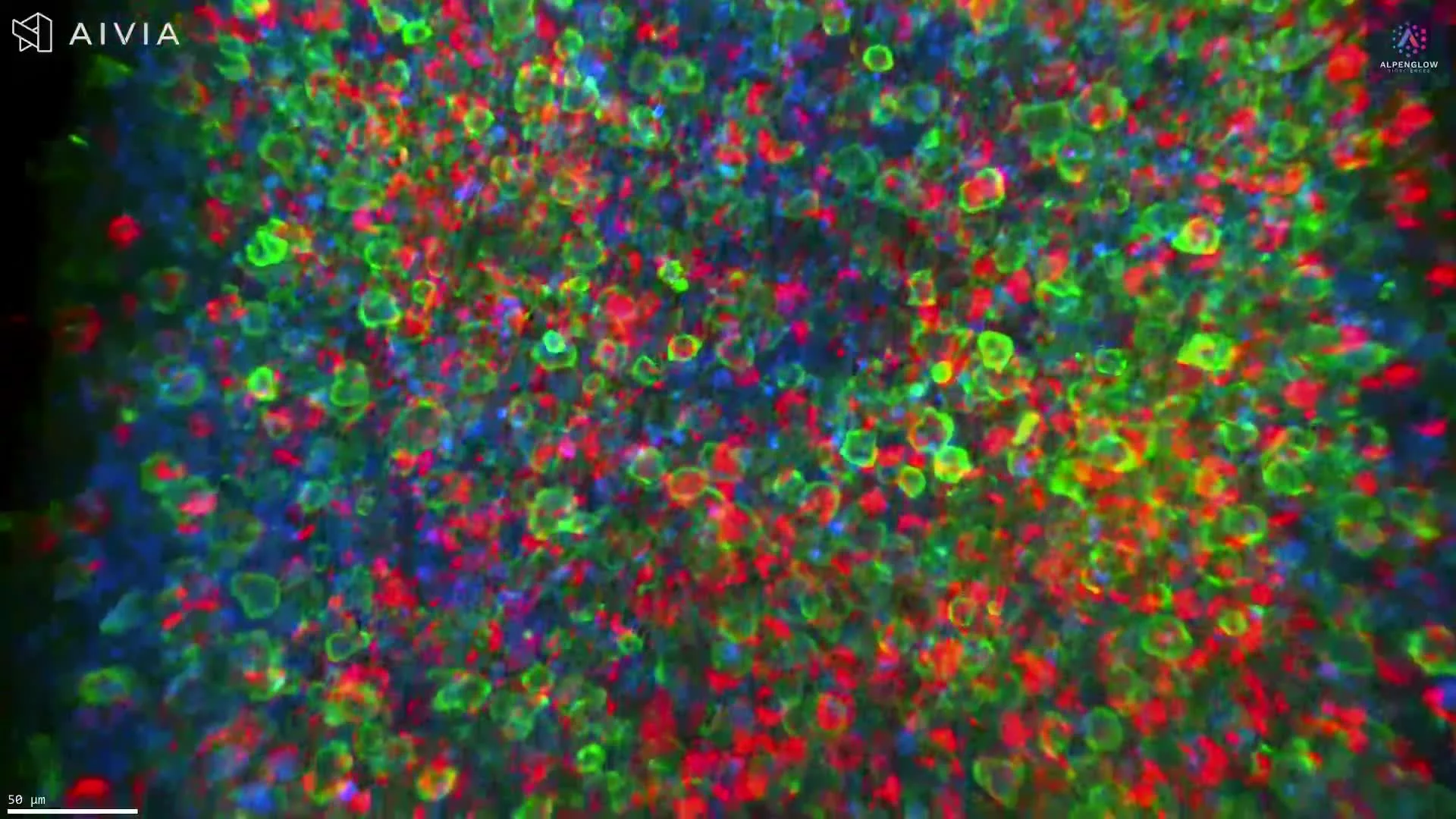
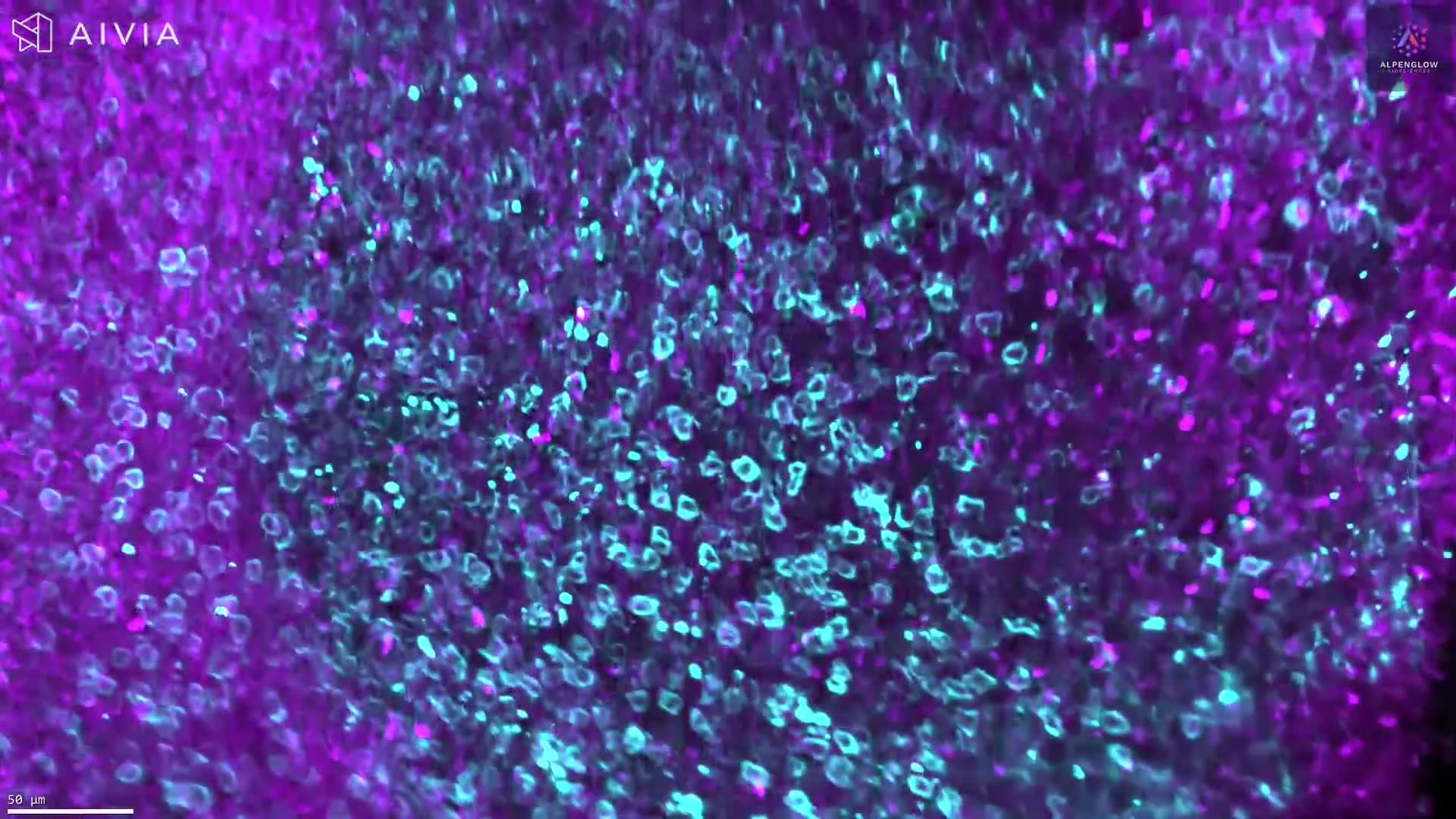
See 3D immuno-oncology imaging applied to real tumor datasets
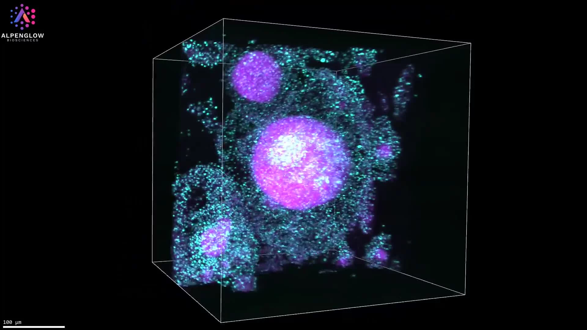
Explore examples of tumor immune microenvironment imaging, including CD45RO memory T cell identification, CD8 spatial mapping, tumor region context, and 3D analysis of immune cells across intact tissue.


Who it supports
Built for teams studying tumor immunity in spatial context
The 3D I/O Pro™ Assay supports immuno-oncology programs that need 3D tissue imaging, spatial profiling, and quantitative tissue analysis across intact tumor samples.
Translational research teams
Study immune infiltration, tumor immune organization, lymphocyte distribution, collagen structure, and tumor microenvironment architecture in full sample context.
Immuno-oncology drug programs
Compare treatment effects across intact tumor samples using volumetric datasets, spatial profiling, and quantitative analysis.
Academic immuno-oncology labs
Explore tumor architecture, immune organization, stromal structure, lymphocyte spatial mapping, and 3D digital pathology readouts.
Use the assay as a service or as part of an in-house Aurora 3D™ workflow. Alpenglow supports immuno-oncology studies from intact tumor tissue preparation through 3D imaging, data processing, and AI-powered analysis.
Start an immuno-oncology study
Ready to see tumor immunity in full 3D context?
Use Alpenglow’s 3D tissue imaging, digital pathology workflows, and AI-powered analysis to study immune exclusion, lymphocyte spatial mapping, TLS organization, collagen structure, and tumor microenvironment architecture across intact tumor samples.