

Revolutionizing Spatial Biology: Advanced 3D Imaging Video Showcase
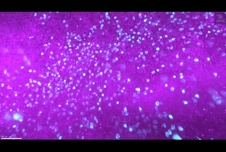
3D Imaging of Human Tonsil Tissue Highlighting Mast Cells with Tryptase Staining
3D view of human tonsil tissue stained with YO-PRO-1 and Tryptase, revealing mast cell distribution and spatial organization in unprecedented detail.

Exploring Prostate Organoids in 3D with LUMI
LUMI revolutionizes 3D imaging of prostate organoids with a seamless workflow: fast low-resolution pre-scans, precise ROI selection on full 3D raw data, and high-resolution multi-well scanning. See how TO-PRO-3 and Eosin staining reveal every detail in this advanced imaging process.
Introducing LUMI: Smarter 3D Imaging
LUMI revolutionizes 3D imaging with an intuitive interface for the HOTLS microscope. Experience effortless scan setup, real-time feedback, and smart microscopy for faster, high-quality results.
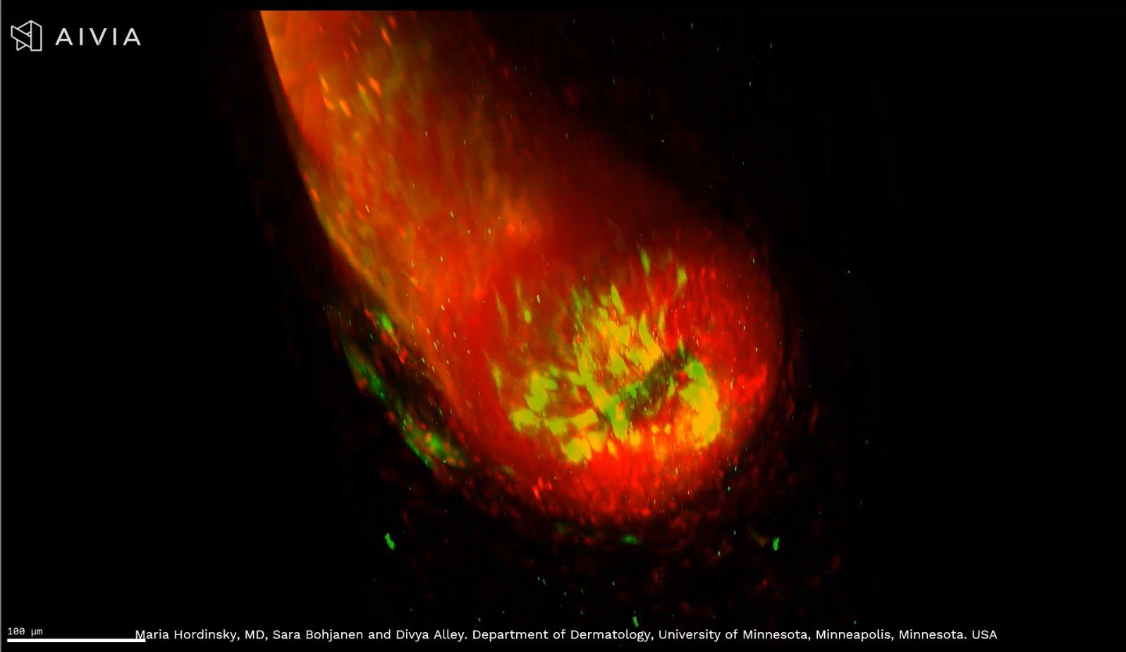
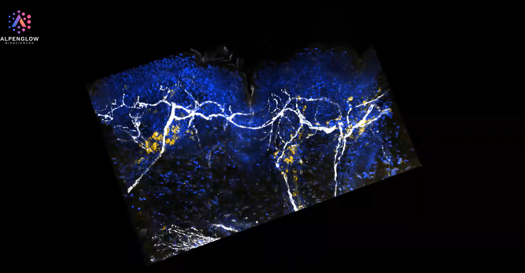
Unlocking the Secrets of Hair Follicles with 3D Imaging
See a hair follicle in stunning 3D detail, from the bulb to the dermal papilla. Using PGP9.5 and TO-PRO-3 staining, this advanced imaging highlights sensory nerve fibers and nuclei, transforming hair research and alopecia areata detection.
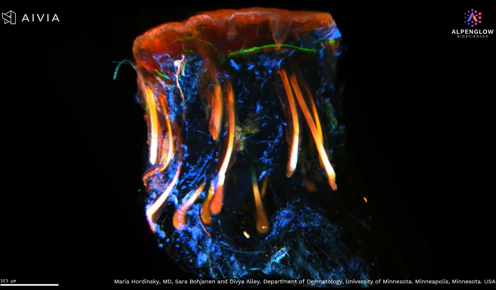
3D Imaging of Scalp Tissue
3D imaging transforms dermatological research by revealing intricate scalp tissue structures with TO-PRO-3, CD45, and PGP9.5 staining. See the difference between full 3D visualization and traditional 2D slices, unlocking new insights into atopic dermatitis, alopecia areata, and more.
Unprecedented Detail in Atopic Dermatitis with 3D Imaging
Experience the power of 3D imaging in dermatology with this high-resolution video of an atopic dermatitis skin biopsy. Visualizing nerves (PGP9.5), immune cells (CD45), and nuclear structures (TOPRO3) in true spatial context, this technology reveals details impossible to capture with traditional tissue sections—offering unprecedented clarity and quantifiable insights for research.
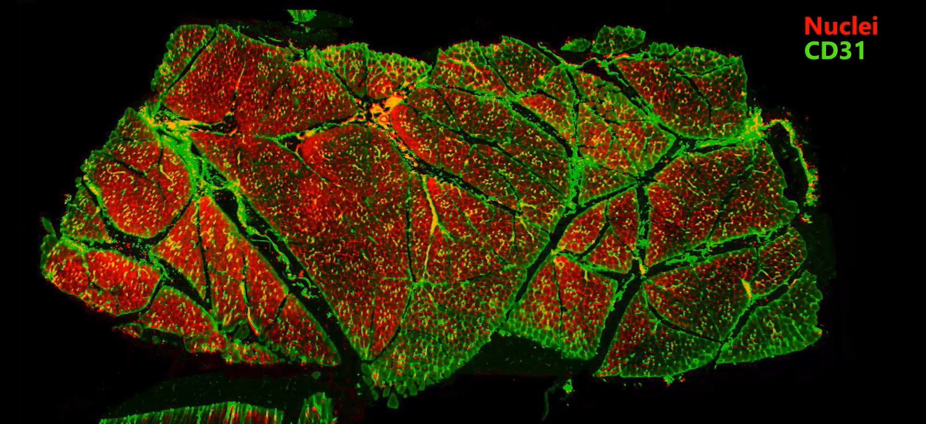
2 mm pig muscle section stained with CD31 and YO-PRO-1
2 mm pig muscle section stained with CD31 (green) for endothelial cells and YO-PRO-1 (red) for nuclei, revealing the intricate vascular network in extraordinary detail.
Using our Hybrid-Open-Top Light Sheet microscope, the 3Di, we seamlessly transition from fast, low-resolution overviews to high-resolution imaging of key regions. This unique approach delivers unparalleled speed and precision, uncovering insights impossible with 2D methods.
Skin biopsy in 3D: from low to high resolution
Journey through the delicate nerve structures of the epidermis and dermis at low resolution, and zoom in for an awe-inspiring close-up of lymphocyte distribution around these dynamic networks. This visualization not only captures the stunning complexity of skin biology but also delivers actionable insights, propelling advancements in dermatology research.
CD45 staining in whole skin biopsy
CD45 staining in whole skin biopsy captured through advanced 3D imaging.
Explore the dynamic interplay of lymphocyte distribution around the nerve network and the complex cellular architecture of the skin.
Appreciate the result of precise data management and image segmentation that unveils lymphocyte distribution around nerves with remarkable accuracy.
Lymphocytes Cluster Near Nerves in Atopic Dermatitis
Delve into the intricate details of a lesional atopic dermatitis sample captured with cutting-edge 40X high-resolution 3D imaging. The sample is vividly stained with TO-PRO-3 blue for nuclei, PGP 9.5 white for nerves, and CD45 yellow for lymphocytes. Marvel at the precise preservation of epidermal and dermal structures and the clear visualization of immune-cell clusters in close proximity to specific nerve fibers.

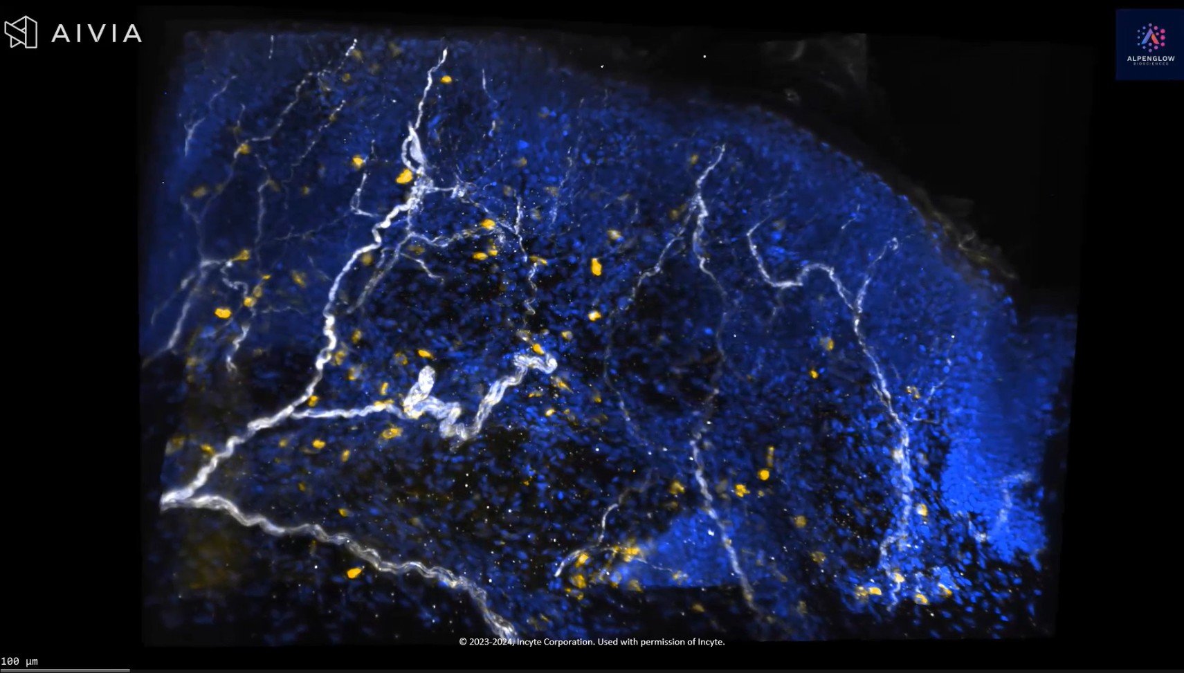
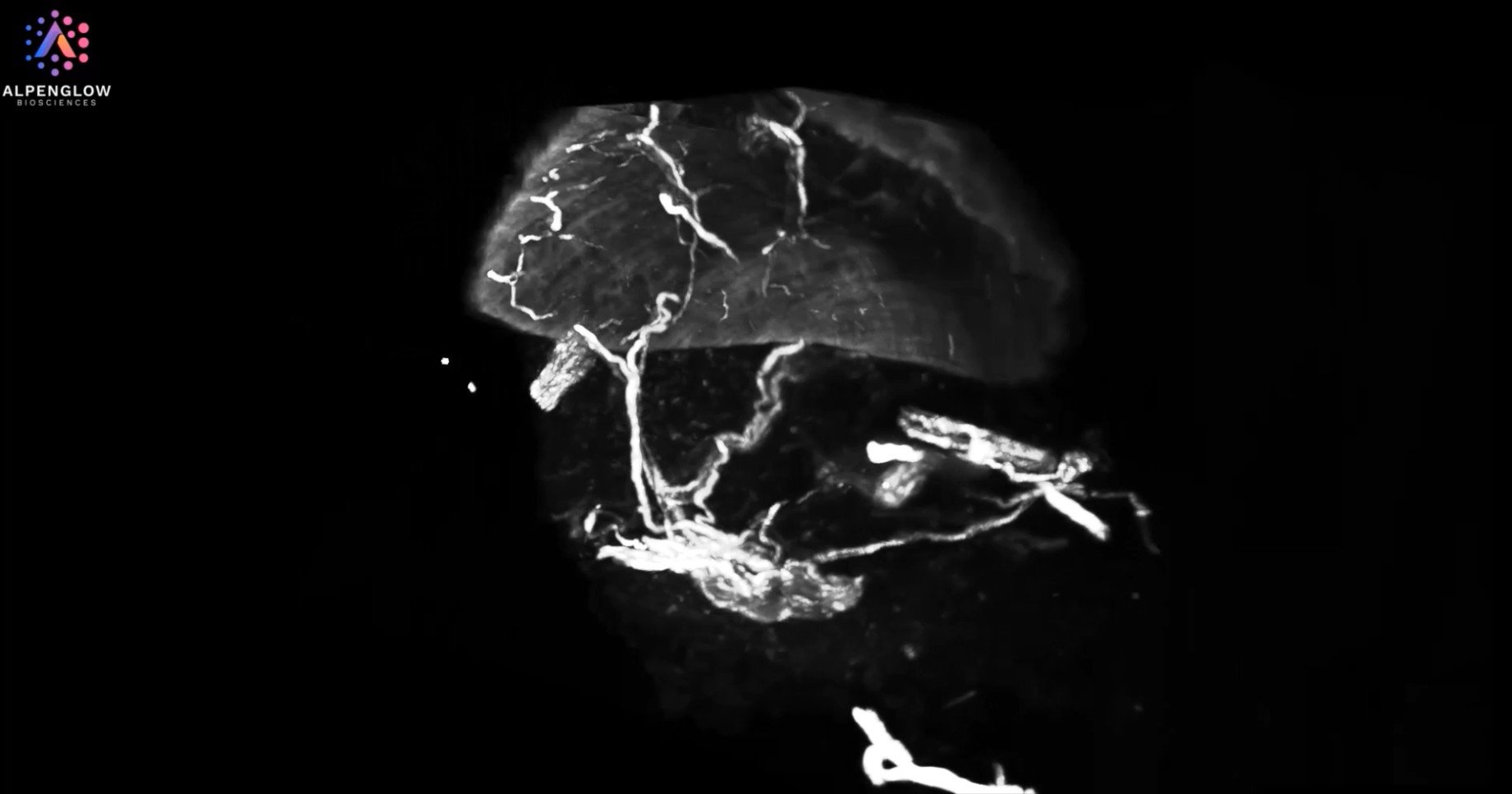
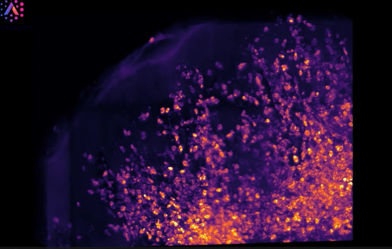
3D Imaging of Glioblastoma in Whole Mouse Brain
3D imaging of a glioblastoma in a mouse brain, from whole-organ visualization to high-resolution tumor analysis.
3D view of Human Normal Adjacent Colorectal Cancer
Explore this cutting-edge 3D imaging of Human Normal Adjacent Colorectal Cancer
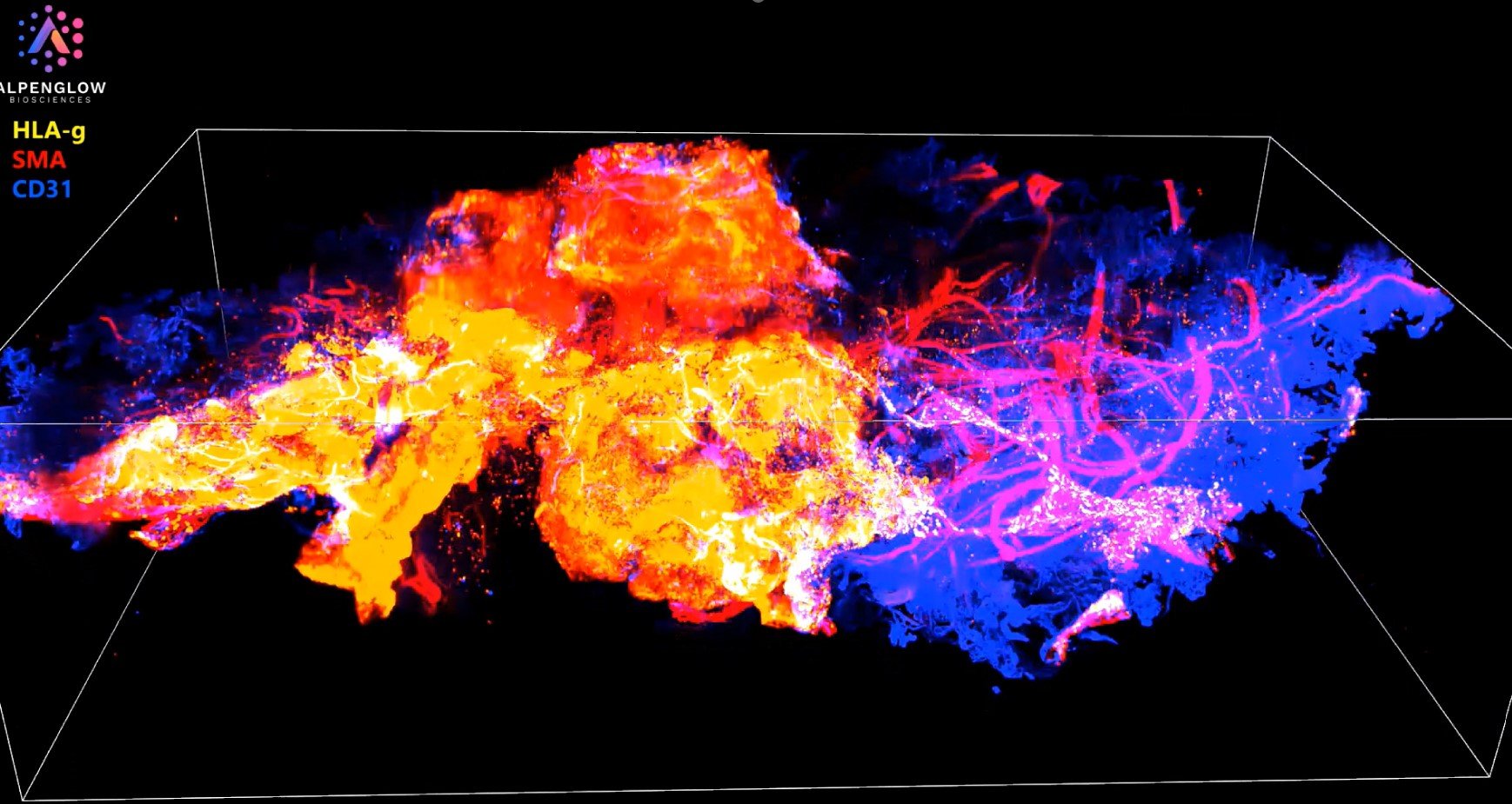
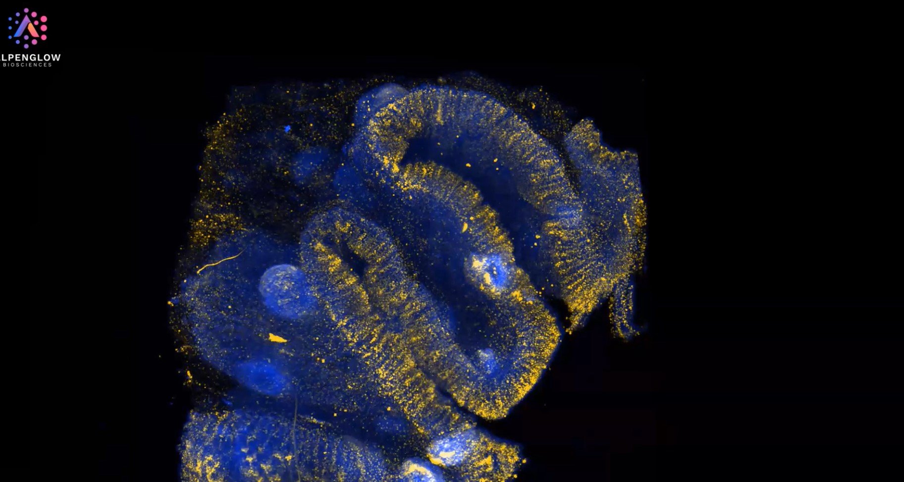
Exploring Human Placenta in 3D: A New Era in Spatial Biology
Experience the intricate architecture of human placenta tissue in this captivating 3D video, designed to showcase the power of advanced spatial biology.

Whole murine heart staining and imaging
This video captures 3D imaging of a whole murine heart, highlighting the complexity and detail of vascular structures. Smooth muscle actin (SMA) stains the blood vessels in green, providing a vivid view of their architecture, while mural cells are highlighted in purple.
Identification of B Cells and Follicular Dendritic Cells through CD21 Staining
Low resolution image of human tonsil measuring up to 3mm in x,y,z, stained with Biocare Bu32 anti-CD21. This antibody has been optimized for a variety of human tissue including FFPE samples.
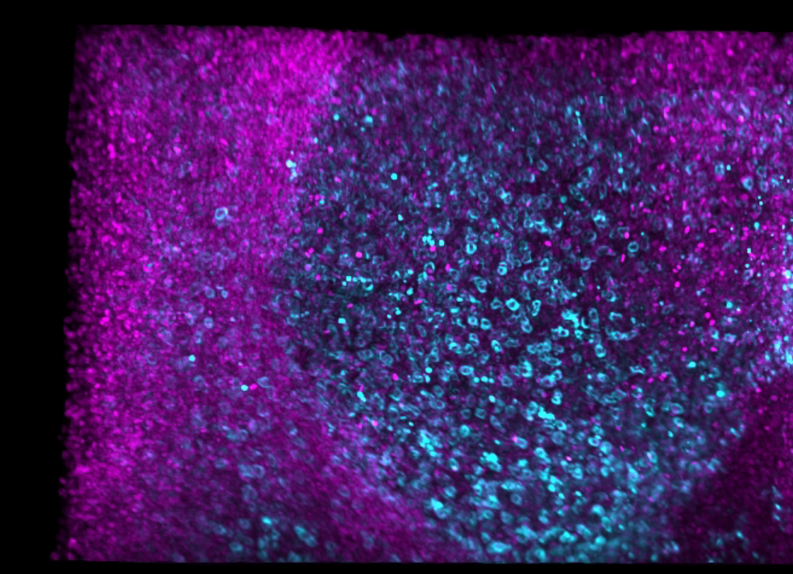
Identification of Memory T-Cells through CD45RO Staining
High resolution image of human tonsil measuring up to 3mm in x,y,z, stained with Biolegend anti-CD45RO. This antibody has been optimized for a variety of human tissue including FFPE samples.
Deep Tissue Staining with anti-CD3 to Identify T-cells
Low resolution image of human tonsil measuring up to 3mm in x,y,z, stained with Biocare Medical BC33 anti-CD3. This antibody has been optimized for a variety of human tissue including FFPE samples.
Identifying cytotoxic T-cells in cleared, thick tissue samples through CD8 Staining
Low resolution image of human tonsil measuring up to 3mm in x,y,z, stained with Biocare Medical SP6 anti-CD8. This antibody has been optimized for a variety of human tissue including FFPE samples.
3D Visualization of Cytotoxic T Cells and Macrophages Using CD8 and CD68 Staining
Explore the intricate distribution of immune cells in human tonsil tissue. This 3D visualization highlights cytotoxic T cells (CD8), macrophages (CD68), and nuclei (TO-PRO-3), offering a detailed view of immune architecture.
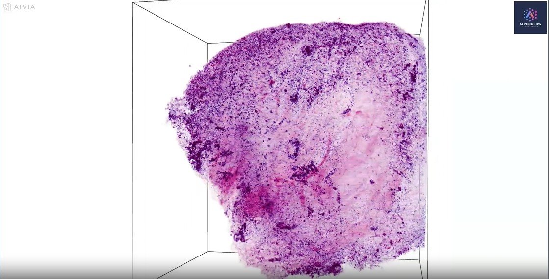
Low-resolution imaging of a human dorsal root ganglion (DRG) sample
This video presents low-resolution (scout) imaging of a human dorsal root ganglion (DRG) sample provided by AnaBios. The sample features computational H&E staining, with ToPro-3 highlighting nuclei and Eosin-capturing protein structures.