
Pathology
REIMAGINED IN 3D

Alpenglow is transforming the field of pathology
Our end-to-end solution includes seamless workflow from sample prep to visualization with 3D analysis. We use an open top light-sheet (OTLS) microscope with patented geometry that allows for ease of use by analyzing entire tissue samples or conventional multi-well plates. This fully automated system facilitates high throughput analysis, while cloud computing and AI-enabled software allows researchers to visualize and quantify complex spatial biology applications in 3D.

3D is a differentiator
Our visualization and analysis allow for a deeper understanding of spatial biology that would not be possible with conventional 2D techniques.

Core applications where 3D is superior to 2D
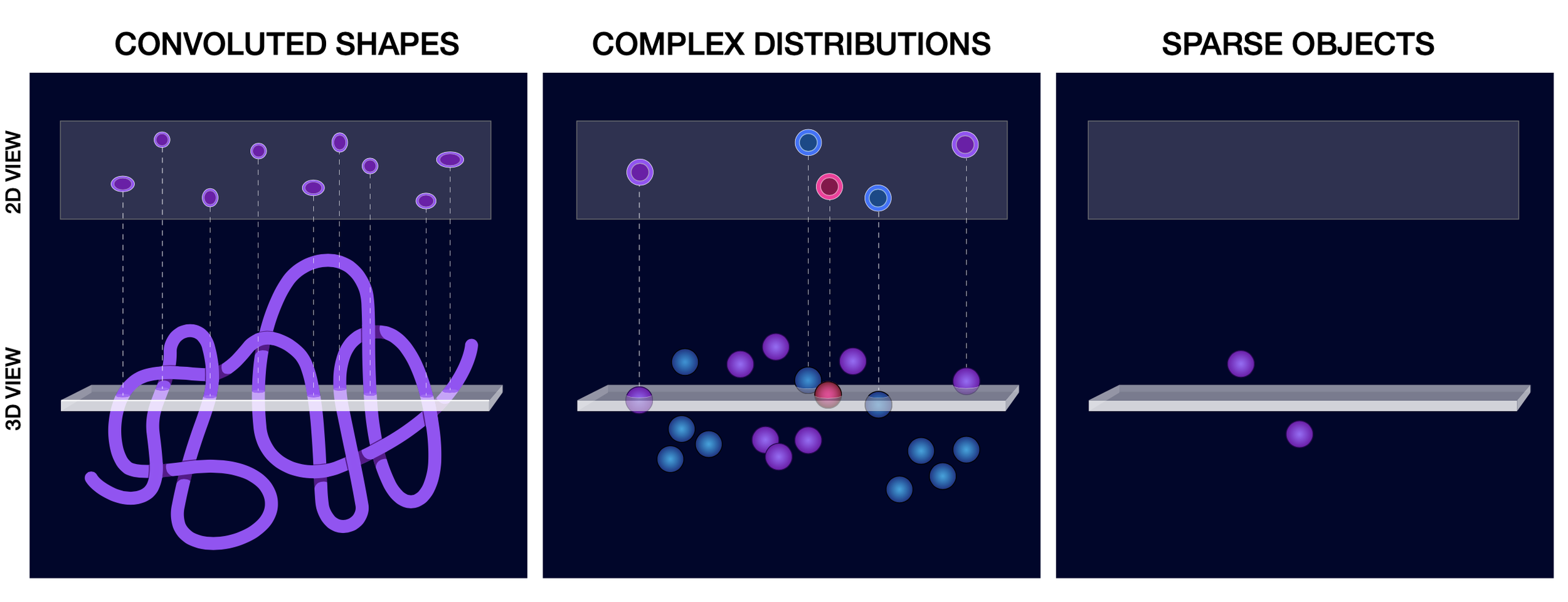
3D Morphology of Convoluted Shapes
Most biologic structures are best quantified in 3D: vasculature, neurons, lymphatics, glands, etc. Alpenglow’s technology provides critical 3D Metrics: volume, total path length, mean diameter, branch count, branch angle, fractal dimension, etc.
Use cases:
· Neuroscience, enteric nervous system, peripheral nervous system
· Vascular changes in dementia, tumors, ischemia, neoangiogensis
· Fibrosis in wide variety of disease including liver, kidney, and lung conditions
Assessment of Complex Cellular Distributions
3D is the best way to understand complex architectures (i.e., microenvironment) in tumors, fibrosis, immune mediated inflammatory disorders, etc.
Use cases:
· Tumor microenvironment
· Amyloid & tau protein distribution
· Immune cell composition / colocalization
Detection of Rare or Sparse Objects
Identify rare cells or drug targets, 2D sectioning will often miss these objects.
Use cases:
· Genetically labeled rare cells
· Biodistribution of drug at target studies
· Stem / progenitor cell studies
· Subclones within PDX models
Doing what is Otherwise Impossible at Scale
Imaging large, unique, or synthetic organs to generate structural maps of large volumes.
Solutions for Drug Discovery, Translational Research & Clinical Diagnostics
Efficacy and dosage analysis
3D spatial biology can provide deep insights into mechanisms of action, drug efficacy, and appropriate dosage. Alpenglow’s technology is particularly well suited for diseases with complex three-dimensional structures like oncology, fibrosis, and neuroscience applications. Using Al-enabled 3D spatial analysis can also give much better quantitative insights into correct drug dosages over traditional, subjective pathologist scoring.
Toxicity and biodistribution analysis
In animal models, imaging an entire tissue sample provides much more insight into possible toxic side effects and the biodistribution of a therapeutic candidate. Catching toxicity with traditional 2D slide-based pathology is prone to sampling errors. Visualizing whole organs using Alpenglow’s 3D spatial biology technology can detect rare toxicity events before compounds reach humans in Phase I clinical trials.
Failed asset screening
During the drug selection process, many viable drug candidates can be cast aside due to incomplete results or lack of efficacy in one domain while having potential applications to other tissues, targets, or disease areas. Alpenglow can rapidly screen biobank samples from previous trials and identify potential efficacious drugs or run simulations for efficacy in other domains. This is achieved by scanning 100% of the tissue and applying Al-enabled 3D deep insights.
Predictive Capabilities
On top of the above offerings, Alpenglow’s proprietary algorithms can be trained to predict molecular alterations, patient outcomes, and patient response to drugs. While these insights can be used in many contexts, they are most helpful in patient selection for clinical trials, developing companion diagnostics, and future therapeutic development and qualification.